 Movie
Movie Controller

[English] 日本語
 Yorodumi
- EMDB-1395: Incorporation of aminoacyl-tRNA into the ribosome as seen by cryo... -
+ Open data

- Basic information
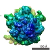
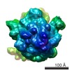
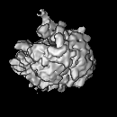
| Entry | Database: EMDB / ID: EMD-1395 | |||||||||
|---|---|---|---|---|---|---|---|---|---|---|
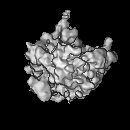
| Title | Incorporation of aminoacyl-tRNA into the ribosome as seen by cryo-electron microscopy. | |||||||||
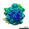
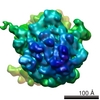
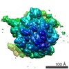
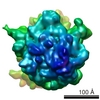
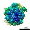
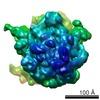
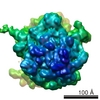
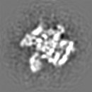
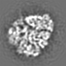
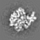
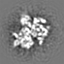
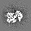
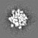
 Map data Map data | Cryo-EM map of E.coli 70S ribosome | |||||||||
 Sample Sample |
| |||||||||
| Biological species |  | |||||||||
| Method | single particle reconstruction / cryo EM / Resolution: 12.8 Å | |||||||||
 Authors Authors | Valle M / Zavialov A / Li W / Stagg SM / Sengupta J / Nielsen RC / Nissen P / Harvey SC / Ehrenberg M / Frank J | |||||||||
 Citation Citation | Journal: Nat Struct Biol / Year: 2003 Title: Incorporation of aminoacyl-tRNA into the ribosome as seen by cryo-electron microscopy. Authors: Mikel Valle / Andrey Zavialov / Wen Li / Scott M Stagg / Jayati Sengupta / Rikke C Nielsen / Poul Nissen / Stephen C Harvey / Måns Ehrenberg / Joachim Frank /  Abstract: Aminoacyl-tRNAs (aa-tRNAs) are delivered to the ribosome as part of the ternary complex of aa-tRNA, elongation factor Tu (EF-Tu) and GTP. Here, we present a cryo-electron microscopy (cryo-EM) study, ...Aminoacyl-tRNAs (aa-tRNAs) are delivered to the ribosome as part of the ternary complex of aa-tRNA, elongation factor Tu (EF-Tu) and GTP. Here, we present a cryo-electron microscopy (cryo-EM) study, at a resolution of approximately 9 A, showing that during the incorporation of the aa-tRNA into the 70S ribosome of Escherichia coli, the flexibility of aa-tRNA allows the initial codon recognition and its accommodation into the ribosomal A site. In addition, a conformational change observed in the GTPase-associated center (GAC) of the ribosomal 50S subunit may provide the mechanism by which the ribosome promotes a relative movement of the aa-tRNA with respect to EF-Tu. This relative rearrangement seems to facilitate codon recognition by the incoming aa-tRNA, and to provide the codon-anticodon recognition-dependent signal for the GTPase activity of EF-Tu. From these new findings we propose a mechanism that can explain the sequence of events during the decoding of mRNA on the ribosome. | |||||||||
| History |
|
- Structure visualization
| Movie |
 Movie viewer Movie viewer |
|---|---|
| Structure viewer | EM map: SurfViewMolmilJmol/JSmol |
| Supplemental images |

- Downloads & links
-EMDB archive
| Map data | emd_1395.map.gz | 7.9 MB | EMDB map data format | |
|---|---|---|---|---|
| Header (meta data) | emd-1395-v30.xmlemd-1395.xml | 8.7 KB 8.7 KB | Display Display | EMDB header |
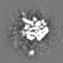
| Images | 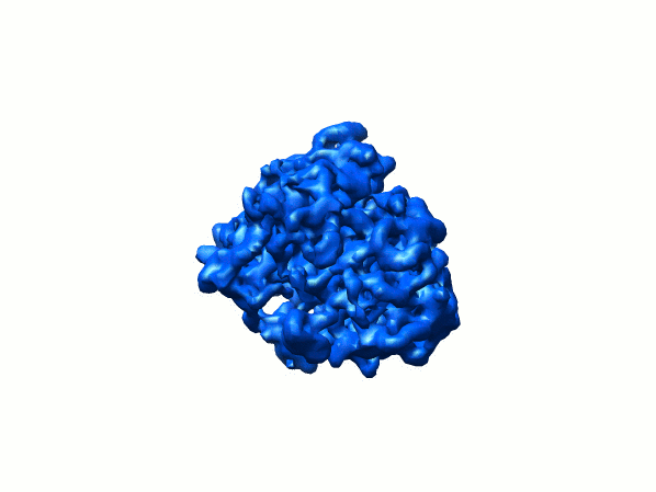 1395.gif 1395.gif | 38.6 KB | ||
| Archive directory |  http://ftp.pdbj.org/pub/emdb/structures/EMD-1395ftp://ftp.pdbj.org/pub/emdb/structures/EMD-1395 http://ftp.pdbj.org/pub/emdb/structures/EMD-1395ftp://ftp.pdbj.org/pub/emdb/structures/EMD-1395 | HTTPS FTP |
-Validation report
| Summary document | emd_1395_validation.pdf.gz | 241.1 KB | Display | EMDB validaton report |
|---|---|---|---|---|
| Full document | emd_1395_full_validation.pdf.gz | 240.2 KB | Display | |
| Data in XML | emd_1395_validation.xml.gz | 6 KB | Display | |
| Arichive directory | https://ftp.pdbj.org/pub/emdb/validation_reports/EMD-1395ftp://ftp.pdbj.org/pub/emdb/validation_reports/EMD-1395 | HTTPS FTP |
-Related structure data
| Related structure data | 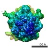 1055C 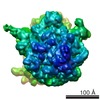 1056C  1qzaC 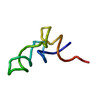 1qzbC 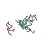 1qzcC 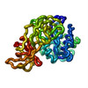 1qzdC 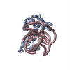 1r2wC  1r2xC C: citing same article ( |
|---|---|
| Similar structure data |
-Links
| EMDB pages | EMDB (EBI/PDBe) / EMDataResource |
|---|---|
| Related items in Molecule of the Month |

-Map
| File | Download / File: emd_1395.map.gz / Format: CCP4 / Size: 8.2 MB / Type: IMAGE STORED AS FLOATING POINT NUMBER (4 BYTES) | ||||||||||||||||||||||||||||||||||||||||||||||||||||||||||||||||||||
|---|---|---|---|---|---|---|---|---|---|---|---|---|---|---|---|---|---|---|---|---|---|---|---|---|---|---|---|---|---|---|---|---|---|---|---|---|---|---|---|---|---|---|---|---|---|---|---|---|---|---|---|---|---|---|---|---|---|---|---|---|---|---|---|---|---|---|---|---|---|
| Annotation | Cryo-EM map of E.coli 70S ribosome | ||||||||||||||||||||||||||||||||||||||||||||||||||||||||||||||||||||
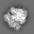
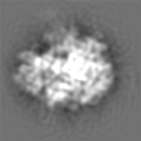
| Projections & slices | Image control
Images are generated by Spider. | ||||||||||||||||||||||||||||||||||||||||||||||||||||||||||||||||||||
| Voxel size | X=Y=Z: 2.82 Å | ||||||||||||||||||||||||||||||||||||||||||||||||||||||||||||||||||||
| Density |
| ||||||||||||||||||||||||||||||||||||||||||||||||||||||||||||||||||||
| Symmetry | Space group: 1 | ||||||||||||||||||||||||||||||||||||||||||||||||||||||||||||||||||||
| Details | EMDB XML:
CCP4 map header:
| ||||||||||||||||||||||||||||||||||||||||||||||||||||||||||||||||||||
 Z (Sec.)
 Y (Row.)
 X (Col.)


-Supplemental data
- Sample components
-Entire : 70S Post-initiation complex with fMettRNAfMet
| Entire | Name: 70S Post-initiation complex with fMettRNAfMet |
|---|---|
| Components |
|
-Supramolecule #1000: 70S Post-initiation complex with fMettRNAfMet
| Supramolecule | Name: 70S Post-initiation complex with fMettRNAfMet / type: sample / ID: 1000 / Number unique components: 3 |
|---|
-Supramolecule #1: Post-initiation Complex
| Supramolecule | Name: Post-initiation Complex / type: complex / ID: 1 / Ribosome-details: ribosome-prokaryote: ALL |
|---|
-Macromolecule #1: tRNA
| Macromolecule | Name: tRNA / type: rna / ID: 1 / Classification: TRANSFER / Structure: DOUBLE HELIX / Synthetic?: No |
|---|---|
| Source (natural) | Organism: |
-Macromolecule #2: mRNA
| Macromolecule | Name: mRNA / type: rna / ID: 2 / Classification: OTHER / Structure: SINGLE STRANDED / Synthetic?: No |
|---|---|
| Source (natural) | Organism: |
-Experimental details
-Structure determination
| Method | cryo EM |
|---|---|
 Processing Processing | single particle reconstruction |
| Aggregation state | particle |
-Sample preparation
| Vitrification | Cryogen name: ETHANE / Details: Rapid-freezing in liquid ethane |
|---|
- Electron microscopy
| Microscope | FEI TECNAI F20 |
|---|---|
| Temperature | Average: 93 K |
| Alignment procedure | Legacy - Electron beam tilt params: 0 |
| Image recording | Category: FILM / Film or detector model: KODAK SO-163 FILM / Digitization - Scanner: ZEISS SCAI / Digitization - Sampling interval: 14 µm |
| Tilt angle min | 0 |
| Tilt angle max | 0 |
| Electron beam | Acceleration voltage: 200 kV / Electron source:  FIELD EMISSION GUN FIELD EMISSION GUN |
| Electron optics | Calibrated magnification: 49650 / Illumination mode: FLOOD BEAM / Imaging mode: BRIGHT FIELD / Nominal defocus max: 4.0 µm / Nominal defocus min: 2.0 µm / Nominal magnification: 50000 |
| Sample stage | Specimen holder: cryo transfer / Specimen holder model: GATAN LIQUID NITROGEN |
| Experimental equipment |  Model: Tecnai F20 / Image courtesy: FEI Company |
-Image processing
| CTF correction | Details: CTF correctionn of 3D map |
|---|---|
| Final reconstruction | Applied symmetry - Point group: C1 (asymmetric) / Algorithm: OTHER / Resolution.type: BY AUTHOR / Resolution: 12.8 Å / Resolution method: FSC 0.5 CUT-OFF / Software - Name: SPIDER package |
