Movie
Movie Controller

[English] 日本語
 Yorodumi
- EMDB-5680: Cryo-electron microscopy of a trimeric soluble HIV Env construct,... -
+ Open data

- Basic information
| Entry | Database: EMDB / ID: EMD-5680 | |||||||||
|---|---|---|---|---|---|---|---|---|---|---|
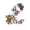
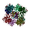
| Title | Cryo-electron microscopy of a trimeric soluble HIV Env construct, gp140 SOSIP, in complex with Fab Z13e1 | |||||||||
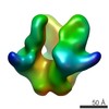
 Map data Map data | Reconstruction of trimeric HIV gp140 with MPER FAB | |||||||||
 Sample Sample |
| |||||||||
 Keywords Keywords | HIV / gp140 / SOSIP / Z13e1 | |||||||||
| Biological species |   Human immunodeficiency virus 1 / unidentified (others) Human immunodeficiency virus 1 / unidentified (others) | |||||||||
| Method | single particle reconstruction / cryo EM / Resolution: 18.5 Å | |||||||||
 Authors Authors | Harris AK / Bartesaghi A / Milne JL / Subramaniam S | |||||||||
 Citation Citation | Journal: J Virol / Year: 2013 Title: HIV-1 envelope glycoprotein trimers display open quaternary conformation when bound to the gp41 membrane-proximal external-region-directed broadly neutralizing antibody Z13e1. Authors: Audray K Harris / Alberto Bartesaghi / Jacqueline L S Milne / Sriram Subramaniam /  Abstract: We describe cryo-electron microscopic studies of the interaction between the ectodomain of the trimeric HIV-1 envelope glycoprotein (Env) and Z13e1, a broadly neutralizing antibody that targets the ...We describe cryo-electron microscopic studies of the interaction between the ectodomain of the trimeric HIV-1 envelope glycoprotein (Env) and Z13e1, a broadly neutralizing antibody that targets the membrane-proximal external region (MPER) of the gp41 subunit. We show that Z13e1-bound Env displays an open quaternary conformation similar to the CD4-bound conformation. Our results support the idea that MPER-directed antibodies, such as Z13e1, block viral entry by interacting with Env at a step after CD4 activation. | |||||||||
| History |
|
- Structure visualization
| Movie |
 Movie viewer Movie viewer |
|---|---|
| Structure viewer | EM map: SurfViewMolmilJmol/JSmol |
| Supplemental images |

- Downloads & links
-EMDB archive
| Map data | emd_5680.map.gz | 5.2 MB | EMDB map data format | |
|---|---|---|---|---|
| Header (meta data) | emd-5680-v30.xmlemd-5680.xml | 9.9 KB 9.9 KB | Display Display | EMDB header |
| Images | 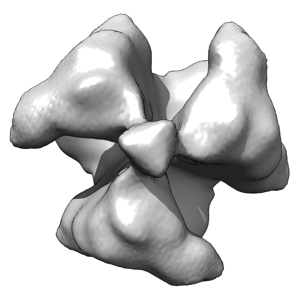 emd_5680_1.jpg emd_5680_1.jpg | 82 KB | ||
| Archive directory |  http://ftp.pdbj.org/pub/emdb/structures/EMD-5680ftp://ftp.pdbj.org/pub/emdb/structures/EMD-5680 http://ftp.pdbj.org/pub/emdb/structures/EMD-5680ftp://ftp.pdbj.org/pub/emdb/structures/EMD-5680 | HTTPS FTP |
-Validation report
| Summary document | emd_5680_validation.pdf.gz | 78.7 KB | Display | EMDB validaton report |
|---|---|---|---|---|
| Full document | emd_5680_full_validation.pdf.gz | 77.8 KB | Display | |
| Data in XML | emd_5680_validation.xml.gz | 494 B | Display | |
| Arichive directory | https://ftp.pdbj.org/pub/emdb/validation_reports/EMD-5680ftp://ftp.pdbj.org/pub/emdb/validation_reports/EMD-5680 | HTTPS FTP |
-Related structure data
| Similar structure data |
|---|


-Links
| EMDB pages | EMDB (EBI/PDBe) / EMDataResource |
|---|
-Map
| File | Download / File: emd_5680.map.gz / Format: CCP4 / Size: 23.2 MB / Type: IMAGE STORED AS FLOATING POINT NUMBER (4 BYTES) | ||||||||||||||||||||||||||||||||||||||||||||||||||||||||||||||||||||
|---|---|---|---|---|---|---|---|---|---|---|---|---|---|---|---|---|---|---|---|---|---|---|---|---|---|---|---|---|---|---|---|---|---|---|---|---|---|---|---|---|---|---|---|---|---|---|---|---|---|---|---|---|---|---|---|---|---|---|---|---|---|---|---|---|---|---|---|---|---|
| Annotation | Reconstruction of trimeric HIV gp140 with MPER FAB | ||||||||||||||||||||||||||||||||||||||||||||||||||||||||||||||||||||
| Voxel size | X=Y=Z: 1.35 Å | ||||||||||||||||||||||||||||||||||||||||||||||||||||||||||||||||||||
| Density |
| ||||||||||||||||||||||||||||||||||||||||||||||||||||||||||||||||||||
| Symmetry | Space group: 1 | ||||||||||||||||||||||||||||||||||||||||||||||||||||||||||||||||||||
| Details | EMDB XML:
CCP4 map header:
| ||||||||||||||||||||||||||||||||||||||||||||||||||||||||||||||||||||
-Supplemental data
- Sample components
-Entire : Molecular structure of KNH1144 SOSIP gp140 with Z13e1 Fab
| Entire | Name: Molecular structure of KNH1144 SOSIP gp140 with Z13e1 Fab |
|---|---|
| Components |
|
-Supramolecule #1000: Molecular structure of KNH1144 SOSIP gp140 with Z13e1 Fab
| Supramolecule | Name: Molecular structure of KNH1144 SOSIP gp140 with Z13e1 Fab type: sample / ID: 1000 / Oligomeric state: trimer / Number unique components: 2 |
|---|
-Macromolecule #1: Envelope glycoprotein
| Macromolecule | Name: Envelope glycoprotein / type: protein_or_peptide / ID: 1 / Name.synonym: Env / Number of copies: 3 / Oligomeric state: trimer / Recombinant expression: Yes |
|---|---|
| Source (natural) | Organism: Human immunodeficiency virus 1 / Strain: isolate 00KE_KNH1144 / synonym: HIV-1 |
| Molecular weight | Experimental: 420 KDa |
| Recombinant expression | Organism:  Homo sapiens (human) / Recombinant plasmid: SOSIP-PPI4 and furin-pcDNA3.1 Homo sapiens (human) / Recombinant plasmid: SOSIP-PPI4 and furin-pcDNA3.1 |
-Macromolecule #2: Fab portion of monoclonal antibody Z13e1
| Macromolecule | Name: Fab portion of monoclonal antibody Z13e1 / type: protein_or_peptide / ID: 2 / Name.synonym: Z13e1 Fab / Details: Fab fragment / Recombinant expression: No / Database: NCBI |
|---|---|
| Source (natural) | Organism: unidentified (others) |
| Molecular weight | Theoretical: 50 KDa |
-Experimental details
-Structure determination
| Method | cryo EM |
|---|---|
 Processing Processing | single particle reconstruction |
| Aggregation state | particle |
-Sample preparation
| Concentration | 0.42 mg/mL |
|---|---|
| Buffer | pH: 7.5 / Details: TNE Buffer (10 mM Tris, 150 mM NaCl, 1 mM EDTA) |
| Grid | Details: Quantifoil, plasma cleaned |
| Vitrification | Cryogen name: ETHANE / Chamber humidity: 100 % / Instrument: FEI VITROBOT MARK III Method: Blot for 6 seconds (blot offset -2) and plunge into an ethane slurry cooled by liquid nitrogen. |
- Electron microscopy
| Microscope | FEI TITAN KRIOS |
|---|---|
| Date | Aug 31, 2009 |
| Image recording | Category: CCD / Film or detector model: GATAN ULTRASCAN 4000 (4k x 4k) / Number real images: 583 / Average electron dose: 20 e/Å2 |
| Electron beam | Acceleration voltage: 80 kV / Electron source:  FIELD EMISSION GUN FIELD EMISSION GUN |
| Electron optics | Illumination mode: FLOOD BEAM / Imaging mode: BRIGHT FIELD / Nominal defocus max: 2.9 µm / Nominal defocus min: 1.3 µm / Nominal magnification: 59500 |
| Sample stage | Specimen holder model: FEI TITAN KRIOS AUTOGRID HOLDER |
| Experimental equipment |  Model: Titan Krios / Image courtesy: FEI Company |
-Image processing
| Final reconstruction | Resolution.type: BY AUTHOR / Resolution: 18.5 Å / Resolution method: FSC 0.5 CUT-OFF / Software - Name: EMAN1 / Number images used: 7050 |
|---|
-Atomic model buiding 1
| Initial model | PDB ID: |
|---|---|
| Software | Name: Chimera |
| Details | Protocol: Rigid body. Automated fitting procedures |
| Refinement | Space: REAL / Protocol: RIGID BODY FIT |