 Movie
Movie Controller


+ Open data

- Basic information
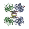
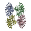
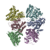
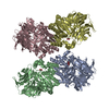
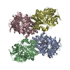
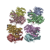
| Entry | Database: PDB / ID: 7asd | |||||||||
|---|---|---|---|---|---|---|---|---|---|---|
| Title | Structure of native royal jelly filaments | |||||||||
 Components Components |
| |||||||||
 Keywords Keywords | PROTEIN FIBRIL /  protein filament / lipoprotein / glycosylation / royal jelly / major royal jelly protein / honeybee protein filament / lipoprotein / glycosylation / royal jelly / major royal jelly protein / honeybee | |||||||||
| Function / homology |  Function and homology information Function and homology informationcaste determination, influence by environmental factors / defense response to fungus / killing of cells of another organism / defense response to Gram-negative bacterium / defense response to Gram-positive bacterium / extracellular region Similarity search - Function | |||||||||
| Biological species |  Apis mellifera (honey bee) Apis mellifera (honey bee) | |||||||||
| Method | ELECTRON MICROSCOPY / helical reconstruction / cryo EM / Resolution: 3.5 Å | |||||||||
 Authors Authors | Mattei, S. / Ban, A. / Picenoni, A. / Leibundgut, M. / Glockshuber, R. / Boehringer, D. | |||||||||
| Funding support | European Union, 2items
| |||||||||
 Citation Citation | Journal: Nat Commun / Year: 2020 Title: Structure of native glycolipoprotein filaments in honeybee royal jelly. Authors: Simone Mattei / Arvid Ban / Armin Picenoni / Marc Leibundgut / Rudi Glockshuber / Daniel Boehringer /   Abstract: Royal jelly (RJ) is produced by honeybees (Apis mellifera) as nutrition during larval development. The high viscosity of RJ originates from high concentrations of long lipoprotein filaments that ...Royal jelly (RJ) is produced by honeybees (Apis mellifera) as nutrition during larval development. The high viscosity of RJ originates from high concentrations of long lipoprotein filaments that include the glycosylated major royal jelly protein 1 (MRJP1), the small protein apisimin and insect lipids. Using cryo-electron microscopy we reveal the architecture and the composition of RJ filaments, in which the MRJP1 forms the outer shell of the assembly, surrounding stacked apisimin tetramers harbouring tightly packed lipids in the centre. The structural data rationalize the pH-dependent disassembly of RJ filaments in the gut of the larvae. | |||||||||
| History |
|
- Structure visualization
| Movie |
Movie viewer |
|---|---|
| Structure viewer | Molecule: MolmilJmol/JSmol |
- Downloads & links
-Download
| PDBx/mmCIF format | 7asd.cif.gz | 648.2 KB | Display | PDBx/mmCIF format |
|---|---|---|---|---|
| PDB format | pdb7asd.ent.gz | Display | PDB format | |
| PDBx/mmJSON format | 7asd.json.gz | Tree view | PDBx/mmJSON format | |
| Others |  Other downloads Other downloads |
-Validation report
| Arichive directory | https://data.pdbj.org/pub/pdb/validation_reports/as/7asdftp://data.pdbj.org/pub/pdb/validation_reports/as/7asd | HTTPS FTP |
|---|
-Related structure data
| Related structure data |  11892MC M: map data used to model this data C: citing same article ( |
|---|---|
| Similar structure data |

-Links
 PDBj
PDBj


- Assembly
| Deposited unit | 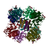
|
|---|---|
| 1 | 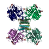
|
| 2 | 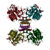
|
-Components
-Protein , 2 types, 16 molecules AABACADAEAFAGAHAABBBCBDBEBFBGBHB
| #1: Protein | Mass: 48934.898 Da / Num. of mol.: 8 / Source method: isolated from a natural source / Source: (natural) Apis mellifera (honey bee) / References: UniProt: O18330#2: Protein | Mass: 7949.325 Da / Num. of mol.: 8 / Source method: isolated from a natural source / Source: (natural) Apis mellifera (honey bee) / Plasmid details: royal jelly / References: UniProt: Q8ISL8 |
|---|
-Sugars , 3 types, 24 molecules 
| #3: Polysaccharide | 2-acetamido-2-deoxy-beta-D-glucopyranose-(1-4)-2-acetamido-2-deoxy-beta-D-glucopyranose / Mass: 424.401 Da / Num. of mol.: 8Source method: isolated from a genetically manipulated source #4: Polysaccharide | beta-D-mannopyranose-(1-4)-2-acetamido-2-deoxy-beta-D-glucopyranose-(1-4)-2-acetamido-2-deoxy-beta- ...beta-D-mannopyranose-(1-4)-2-acetamido-2-deoxy-beta-D-glucopyranose-(1-4)-2-acetamido-2-deoxy-beta-D-glucopyranose / Mass: 586.542 Da / Num. of mol.: 8Source method: isolated from a genetically manipulated source #6: Sugar | ChemComp-NAG / N-Acetylglucosamine Type: D-saccharide, beta linking / Mass: 221.208 Da / Num. of mol.: 8 / Source method: obtained synthetically / Formula: C8H15NO6 Type: D-saccharide, beta linking / Mass: 221.208 Da / Num. of mol.: 8 / Source method: obtained synthetically / Formula: C8H15NO6 |
|---|
-Non-polymers , 2 types, 24 molecules 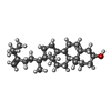

| #5: Chemical | ChemComp-94R / (  Mass: 398.664 Da / Num. of mol.: 16 / Source method: obtained synthetically / Formula: C28H46O Mass: 398.664 Da / Num. of mol.: 16 / Source method: obtained synthetically / Formula: C28H46O#7: Chemical | ChemComp-SO4 / Sulfate Mass: 96.063 Da / Num. of mol.: 8 / Source method: obtained synthetically / Formula: SO4 Mass: 96.063 Da / Num. of mol.: 8 / Source method: obtained synthetically / Formula: SO4 |
|---|
-Details
| Has ligand of interest | N |
|---|
-Experimental details
-Experiment
| Experiment | Method: ELECTRON MICROSCOPY |
|---|---|
| EM experiment | Aggregation state: FILAMENT / 3D reconstruction method: helical reconstruction |
- Sample preparation
| Component | Name: native royal jelly filaments / Type: COMPLEX / Entity ID: #1-#2 / Source: NATURAL |
|---|---|
| Source (natural) | Organism: Apis mellifera (honey bee) |
| Buffer solution | pH: 4 |
| Specimen | Embedding applied: NO / Shadowing applied: NO / Staining applied: NO / Vitrification applied: YES |
| Specimen support | Grid material: COPPER / Grid mesh size: 300 divisions/in. / Grid type: Quantifoil R2/2 |
| Vitrification | Cryogen name: ETHANE-PROPANE |
- Electron microscopy imaging
| Experimental equipment |  Model: Titan Krios / Image courtesy: FEI Company |
|---|---|
| Microscopy | Model: FEI TITAN KRIOS |
| Electron gun | Electron source: FIELD EMISSION GUN / Accelerating voltage: 300 kV / Illumination mode: FLOOD BEAM |
| Electron lens | Mode: BRIGHT FIELDBright-field microscopy / Nominal magnification: 105000 X / Calibrated magnification: 119050 X / Cs: 2.7 mm / C2 aperture diameter: 100 µm |
| Image recording | Average exposure time: 1.7 sec. / Electron dose: 82 e/Å2 / Film or detector model: GATAN K3 BIOQUANTUM (6k x 4k) |
| EM imaging optics | Energyfilter name: GIF Bioquantum / Energyfilter slit width: 20 eV |
- Processing
| EM software |
| ||||||||||||||||||||||||
|---|---|---|---|---|---|---|---|---|---|---|---|---|---|---|---|---|---|---|---|---|---|---|---|---|---|
| CTF correction | Type: PHASE FLIPPING AND AMPLITUDE CORRECTION | ||||||||||||||||||||||||
| Helical symmerty | Angular rotation/subunit: 64 ° / Axial rise/subunit: 54 Å / Axial symmetry: D2 | ||||||||||||||||||||||||
| 3D reconstruction | Resolution: 3.5 Å / Resolution method: FSC 0.143 CUT-OFF / Num. of particles: 240483 / Algorithm: FOURIER SPACE / Symmetry type: HELICAL |
