 Movie
Movie Controller

[English] 日本語
 Yorodumi
- PDB-3j2i: Structure of late pre-60S ribosomal subunits with nuclear export ... -
+ Open data

- Basic information
| Entry | Database: PDB / ID: 3j2i | ||||||
|---|---|---|---|---|---|---|---|
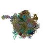
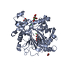
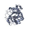
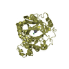
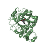
| Title | Structure of late pre-60S ribosomal subunits with nuclear export factor Arx1 bound at the peptide exit tunnel | ||||||
 Components Components |
| ||||||
 Keywords Keywords | RIBOSOMAL PROTEIN / SIGNALING PROTEIN / ribosome / pre-ribosome / cryo-EM | ||||||
| Function / homology |  Function and homology information Function and homology informationmaturation of 5.8S rRNA from tricistronic rRNA transcript (SSU-rRNA, 5.8S rRNA, LSU-rRNA) / maturation of 5.8S rRNA / ribosomal large subunit binding / preribosome, large subunit precursor / ribosomal subunit export from nucleus / translation initiation factor activity / maturation of LSU-rRNA / maturation of LSU-rRNA from tricistronic rRNA transcript (SSU-rRNA, 5.8S rRNA, LSU-rRNA) / assembly of large subunit precursor of preribosome / cytosolic ribosome assembly ...maturation of 5.8S rRNA from tricistronic rRNA transcript (SSU-rRNA, 5.8S rRNA, LSU-rRNA) / maturation of 5.8S rRNA / ribosomal large subunit binding / preribosome, large subunit precursor / ribosomal subunit export from nucleus / translation initiation factor activity / maturation of LSU-rRNA / maturation of LSU-rRNA from tricistronic rRNA transcript (SSU-rRNA, 5.8S rRNA, LSU-rRNA) / assembly of large subunit precursor of preribosome / cytosolic ribosome assembly / ribosomal large subunit biogenesis / positive regulation of cell differentiation / rRNA processing / transcription corepressor activity / azurophil granule lumen / regulation of translation / nucleic acid binding / ribonucleoprotein complex / negative regulation of DNA-templated transcription / ubiquitin protein ligase binding / Neutrophil degranulation / negative regulation of apoptotic process / nucleolus / RNA binding / extracellular exosome / extracellular region / membrane / nucleus / cytoplasm / cytosol Similarity search - Function | ||||||
| Biological species |   Homo sapiens (human) Homo sapiens (human) | ||||||
| Method | ELECTRON MICROSCOPY / single particle reconstruction / cryo EM / Resolution: 11.9 Å | ||||||
 Authors Authors | Bradatsch, B. / Leidig, C. / Granneman, S. / Gnaedig, M. / Tollervey, D. / Boettcher, B. / Beckmann, R. / Hurt, E. | ||||||
 Citation Citation | Journal: Nat Struct Mol Biol / Year: 2012 Title: Structure of the pre-60S ribosomal subunit with nuclear export factor Arx1 bound at the exit tunnel. Authors: Bettina Bradatsch / Christoph Leidig / Sander Granneman / Marén Gnädig / David Tollervey / Bettina Böttcher / Roland Beckmann / Ed Hurt /  Abstract: Preribosomal particles evolve in the nucleus through transient interaction with biogenesis factors before export to the cytoplasm. Here, we report the architecture of the late pre-60S particle, ...Preribosomal particles evolve in the nucleus through transient interaction with biogenesis factors before export to the cytoplasm. Here, we report the architecture of the late pre-60S particle, purified from Saccharomyces cerevisiae, through Arx1, a nuclear export factor with structural homology to methionine aminopeptidases, or its binding partner Alb1. Cryo-EM reconstruction of the Arx1 particle at 11.9-Å resolution reveals regions of extra density on the pre-60S particle attributed to associated biogenesis factors, confirming the immature state of the nascent subunit. One of these densities could be unambiguously assigned to Arx1. Immunoelectron microscopy and UV cross-linking localize Arx1 close to the ribosomal exit tunnel, in direct contact with ES27, a highly dynamic eukaryotic rRNA expansion segment. The binding of Arx1 at the exit tunnel may position this export factor to prevent premature recruitment of ribosome-associated factors active during translation. | ||||||
| History |
|
- Structure visualization
| Movie |
Movie viewer |
|---|---|
| Structure viewer | Molecule: MolmilJmol/JSmol |
- Downloads & links
-Download
| PDBx/mmCIF format | 3j2i.cif.gz | 121.2 KB | Display | PDBx/mmCIF format |
|---|---|---|---|---|
| PDB format | pdb3j2i.ent.gz | 93.7 KB | Display | PDB format |
| PDBx/mmJSON format | 3j2i.json.gz | Tree view | PDBx/mmJSON format | |
| Others |  Other downloads Other downloads |
-Validation report
| Summary document | 3j2i_validation.pdf.gz | 740.2 KB | Display | wwPDB validaton report |
|---|---|---|---|---|
| Full document | 3j2i_full_validation.pdf.gz | 748 KB | Display | |
| Data in XML | 3j2i_validation.xml.gz | 24.4 KB | Display | |
| Data in CIF | 3j2i_validation.cif.gz | 34.8 KB | Display | |
| Arichive directory | https://data.pdbj.org/pub/pdb/validation_reports/j2/3j2iftp://data.pdbj.org/pub/pdb/validation_reports/j2/3j2i | HTTPS FTP |
-Related structure data
| Related structure data | 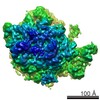 5513MC M: map data used to model this data C: citing same article ( |
|---|---|
| Similar structure data |
-Links
 PDBj
PDBj
- Assembly
| Deposited unit | 
|
|---|---|
| 1 | 
|
| 2 | 
|
-Components
| #1: Protein | Mass: 26476.605 Da / Num. of mol.: 1 / Source method: isolated from a natural source / Source: (natural) |
|---|---|
| #2: Protein | Mass: 43851.879 Da / Num. of mol.: 1 / Source method: isolated from a natural source / Source: (natural) Homo sapiens (human) / References: UniProt: Q9UQ80 |
-Experimental details
-Experiment
| Experiment | Method: ELECTRON MICROSCOPY |
|---|---|
| EM experiment | Aggregation state: PARTICLE / 3D reconstruction method: single particle reconstruction |
- Sample preparation
| Component | Name: pre-60S particle / Type: RIBOSOME / Synonym: pre-60S |
|---|---|
| Molecular weight | Value: 1.3 MDa / Experimental value: NO |
| Buffer solution | pH: 7 Details: 50 mM Tris-HCl, pH 7.5, 100 mM NaCl, 1.5 mM or 5 mM MgCl2, 0.075% (v/v) NP-40 |
| Specimen | Embedding applied: NO / Shadowing applied: NO / Staining applied: NO / Vitrification applied: YES |
| Vitrification | Instrument: FEI VITROBOT MARK IV / Cryogen name: ETHANE / Humidity: 95 % Details: Blot for 2 seconds before plunging in liquid ethane (FEI VITROBOT MARK IV) |
- Electron microscopy imaging
| Experimental equipment |  Model: Titan Krios / Image courtesy: FEI Company |
|---|---|
| Microscopy | Model: FEI TITAN KRIOS / Date: Apr 6, 2012 |
| Electron gun | Electron source:  FIELD EMISSION GUN / Accelerating voltage: 200 kV / Illumination mode: FLOOD BEAM FIELD EMISSION GUN / Accelerating voltage: 200 kV / Illumination mode: FLOOD BEAM |
| Electron lens | Mode: BRIGHT FIELD |
| Specimen holder | Specimen holder model: FEI TITAN KRIOS AUTOGRID HOLDER |
| Image recording | Electron dose: 20 e/Å2 / Film or detector model: GENERIC CCD |
| Radiation | Protocol: SINGLE WAVELENGTH / Monochromatic (M) / Laue (L): M / Scattering type: x-ray |
| Radiation wavelength | Relative weight: 1 |
- Processing
| EM software |
| ||||||||||||
|---|---|---|---|---|---|---|---|---|---|---|---|---|---|
| CTF correction | Details: subvolumes | ||||||||||||
| Symmetry | Point symmetry: C1 (asymmetric) | ||||||||||||
| 3D reconstruction | Method: Projection Matching / Resolution: 11.9 Å / Num. of particles: 100000 / Nominal pixel size: 1.2375 Å / Actual pixel size: 1.2375 Å / Symmetry type: POINT | ||||||||||||
| Refinement step | Cycle: LAST
|
