 Movie
Movie Controller


+ Open data

- Basic information
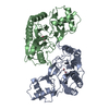
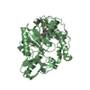
| Entry | Database: PDB / ID: 2v6c | ||||||
|---|---|---|---|---|---|---|---|
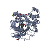
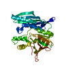
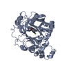
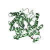
| Title | Crystal structure of ErbB3 binding protein 1 (Ebp1) | ||||||
 Components Components | PROLIFERATION-ASSOCIATED PROTEIN 2G4 | ||||||
 Keywords Keywords |  TRANSCRIPTION REGULATOR / TRANSLATION REGULATION / TRANSLATIONAL REGULATOR / RNA-BINDING / ACETYLATION / RNA BINDING / TRANSCRIPTION / COBALT / NUCLEUS / REPRESSOR / HYDROLASE / CYTOPLASM / TRANSCRIPTION REGULATION / PHOSPHORYLATION / RRNA PROCESSING / RIBONUCLEOPROTEIN TRANSCRIPTION REGULATOR / TRANSLATION REGULATION / TRANSLATIONAL REGULATOR / RNA-BINDING / ACETYLATION / RNA BINDING / TRANSCRIPTION / COBALT / NUCLEUS / REPRESSOR / HYDROLASE / CYTOPLASM / TRANSCRIPTION REGULATION / PHOSPHORYLATION / RRNA PROCESSING / RIBONUCLEOPROTEIN | ||||||
| Function / homology |  Function and homology information Function and homology informationNeutrophil degranulation / positive regulation of cell differentiation / rRNA processing / transcription corepressor activity / regulation of translation / nucleic acid binding / ribonucleoprotein complex / negative regulation of DNA-templated transcription / ubiquitin protein ligase binding / nucleolus ...Neutrophil degranulation / positive regulation of cell differentiation / rRNA processing / transcription corepressor activity / regulation of translation / nucleic acid binding / ribonucleoprotein complex / negative regulation of DNA-templated transcription / ubiquitin protein ligase binding / nucleolus / negative regulation of apoptotic process / RNA binding / nucleus / cytoplasmSimilarity search - Function | ||||||
| Biological species |  MUS MUSCULUS (house mouse) MUS MUSCULUS (house mouse) | ||||||
| Method | X-RAY DIFFRACTION / SYNCHROTRON / MOLECULAR REPLACEMENT / Resolution: 2.5 Å | ||||||
 Authors Authors | Monie, T.P. / Perrin, A.J. / Birtley, J.R. / Curry, S. | ||||||
 Citation Citation | Journal: Embo J. / Year: 2007 Title: Structural Insights Into the Transcriptional and Translational Roles of Ebp1 Authors: Monie, T.P. / Perrin, A.J. / Birtley, J.R. / Sweeney, T.R. / Karakasiliotis, I. / Chaudry, Y. / Roberts, L.O. / Matthews, S. / Goodfellow, I.G. / Curry, S. | ||||||
| History |
| ||||||
| Remark 700 | SHEET THE SHEET STRUCTURE OF THIS MOLECULE IS BIFURCATED. IN ORDER TO REPRESENT THIS FEATURE IN ... SHEET THE SHEET STRUCTURE OF THIS MOLECULE IS BIFURCATED. IN ORDER TO REPRESENT THIS FEATURE IN THE SHEET RECORDS BELOW, TWO SHEETS ARE DEFINED. |
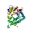
- Structure visualization
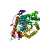
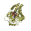
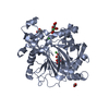
| Structure viewer | Molecule: MolmilJmol/JSmol |
|---|
- Downloads & links
-Download
| PDBx/mmCIF format | 2v6c.cif.gz | 81.8 KB | Display | PDBx/mmCIF format |
|---|---|---|---|---|
| PDB format | pdb2v6c.ent.gz | 60.3 KB | Display | PDB format |
| PDBx/mmJSON format | 2v6c.json.gz | Tree view | PDBx/mmJSON format | |
| Others |  Other downloads Other downloads |
-Validation report
| Arichive directory | https://data.pdbj.org/pub/pdb/validation_reports/v6/2v6cftp://data.pdbj.org/pub/pdb/validation_reports/v6/2v6c | HTTPS FTP |
|---|
-Related structure data

-Links
 PDBj
PDBj


- Assembly
| Deposited unit | 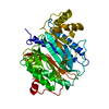
| ||||||||
|---|---|---|---|---|---|---|---|---|---|
| 1 |
| ||||||||
| Unit cell |
|
-Components
| #1: Protein | Mass: 39387.074 Da / Num. of mol.: 1 / Fragment: RESIDUES 7-359 Source method: isolated from a genetically manipulated source Source: (gene. exp.) MUS MUSCULUS (house mouse) / Production host:  ESCHERICHIA COLI (E. coli) / Strain (production host): BL21(DE3) / Variant (production host): ROSETTA (NOVAGEN) / References: UniProt: P50580 ESCHERICHIA COLI (E. coli) / Strain (production host): BL21(DE3) / Variant (production host): ROSETTA (NOVAGEN) / References: UniProt: P50580 | ||
|---|---|---|---|
| #2: Water | ChemComp-HOH / Water Mass: 18.015 Da / Num. of mol.: 55 / Source method: isolated from a natural source / Formula: H2O Mass: 18.015 Da / Num. of mol.: 55 / Source method: isolated from a natural source / Formula: H2O | ||
| Compound details | MAY PLAY A ROLE IN A ERBB3-REGULATED SIGNAL TRANSDUCTI| Sequence details | SEQUENCE CONTAINS 2 MUTATIONS (T279A, K311R) WHICH HAVE BEEN NOTED IN THE LITERATURE AS A PROBABLE ...SEQUENCE CONTAINS 2 MUTATIONS (T279A, K311R) WHICH HAVE BEEN NOTED IN THE LITERATURE | |
-Experimental details
-Experiment
| Experiment | Method: X-RAY DIFFRACTION / Number of used crystals: 1 |
|---|
- Sample preparation
| Crystal | Density Matthews: 2.63 Å3/Da / Density % sol: 53 % / Description: NONE |
|---|---|
| Crystal grow | pH: 7.7 / Details: SEE PAPER, pH 7.7 |
-Data collection
| Diffraction | Mean temperature: 298 K |
|---|---|
| Diffraction source | Source: SYNCHROTRON / Site: EMBL/DESY, HAMBURG  / Beamline: X11 / Wavelength: 0.8123 / Beamline: X11 / Wavelength: 0.8123 |
| Detector | Type: MARRESEARCH / Detector: CCD / Date: Jan 15, 2005 / Details: MIRRORS |
| Radiation | Monochromator: SI CRYSTAL / Protocol: SINGLE WAVELENGTH / Monochromatic (M) / Laue (L): M / Scattering type: x-ray |
| Radiation wavelength | Wavelength: 0.8123 Å / Relative weight: 1 |
| Reflection | Resolution: 2.5→38.4 Å / Num. obs: 14714 / % possible obs: 98.9 % / Observed criterion σ(I): 0 / Redundancy: 3 % / Biso Wilson estimate: 32.9 Å2 / Rmerge(I) obs: 0.08 / Net I/σ(I): 10.4 |
| Reflection shell | Resolution: 2.5→2.66 Å / Redundancy: 3 % / Rmerge(I) obs: 0.41 / Mean I/σ(I) obs: 3.3 / % possible all: 98.9 |
- Processing
| Software |
| ||||||||||||||||||||||||||||||||||||||||||||||||||||||||||||||||||||||||||||||||
|---|---|---|---|---|---|---|---|---|---|---|---|---|---|---|---|---|---|---|---|---|---|---|---|---|---|---|---|---|---|---|---|---|---|---|---|---|---|---|---|---|---|---|---|---|---|---|---|---|---|---|---|---|---|---|---|---|---|---|---|---|---|---|---|---|---|---|---|---|---|---|---|---|---|---|---|---|---|---|---|---|---|
| Refinement | Method to determine structure: MOLECULAR REPLACEMENT Starting model: PDB ENTRIES 1KQ9 AND 1XGS Resolution: 2.5→37.45 Å / Rfactor Rfree error: 0.008 / Data cutoff high absF: 1282714.16 / Isotropic thermal model: RESTRAINED / Cross valid method: THROUGHOUT / σ(F): 0 / Stereochemistry target values: MLF Details: RESIDUES 1-8 AND 361-394 WERE MISSING FROM THE PROTEIN USED IN CRYSALLISATION. RESIDUE 8 WAS MUTATED TO GLU BY THE CLONING PROCEDURE.
| ||||||||||||||||||||||||||||||||||||||||||||||||||||||||||||||||||||||||||||||||
| Solvent computation | Solvent model: FLAT MODEL / Bsol: 21.1966 Å2 / ksol: 0.288707 e/Å3 | ||||||||||||||||||||||||||||||||||||||||||||||||||||||||||||||||||||||||||||||||
| Displacement parameters | Biso mean: 35.2 Å2
| ||||||||||||||||||||||||||||||||||||||||||||||||||||||||||||||||||||||||||||||||
| Refine analyze |
| ||||||||||||||||||||||||||||||||||||||||||||||||||||||||||||||||||||||||||||||||
| Refinement step | Cycle: LAST / Resolution: 2.5→37.45 Å
| ||||||||||||||||||||||||||||||||||||||||||||||||||||||||||||||||||||||||||||||||
| Refine LS restraints |
| ||||||||||||||||||||||||||||||||||||||||||||||||||||||||||||||||||||||||||||||||
| LS refinement shell | Resolution: 2.5→2.66 Å / Rfactor Rfree error: 0.025 / Total num. of bins used: 6
| ||||||||||||||||||||||||||||||||||||||||||||||||||||||||||||||||||||||||||||||||
| Xplor file |
|
