Movie
Movie Controller

[English] 日本語
 Yorodumi
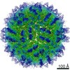
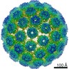
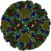
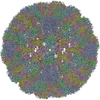
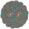
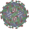
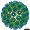
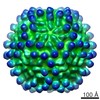
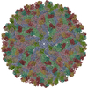
- PDB-3iya: Association of the pr peptides with dengue virus blocks membrane ... -
+ Open data

- Basic information
| Entry | Database: PDB / ID: 3iya | ||||||
|---|---|---|---|---|---|---|---|
| Title | Association of the pr peptides with dengue virus blocks membrane fusion at acidic pH | ||||||
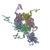
 Components Components |
| ||||||
 Keywords Keywords | VIRUS / prM / E / Icosahedral virus | ||||||
| Function / homology |  Function and homology information Function and homology informationsymbiont-mediated suppression of host JAK-STAT cascade via inhibition of host TYK2 activity / flavivirin / host cell mitochondrion / symbiont-mediated suppression of host JAK-STAT cascade via inhibition of STAT2 activity / symbiont-mediated suppression of host cytoplasmic pattern recognition receptor signaling pathway via inhibition of MAVS activity / viral capsid / double-stranded RNA binding / nucleoside-triphosphate phosphatase / protein complex oligomerization / monoatomic ion channel activity ...symbiont-mediated suppression of host JAK-STAT cascade via inhibition of host TYK2 activity / flavivirin / host cell mitochondrion / symbiont-mediated suppression of host JAK-STAT cascade via inhibition of STAT2 activity / symbiont-mediated suppression of host cytoplasmic pattern recognition receptor signaling pathway via inhibition of MAVS activity / viral capsid / double-stranded RNA binding / nucleoside-triphosphate phosphatase / protein complex oligomerization / monoatomic ion channel activity / mRNA (guanine-N7)-methyltransferase / methyltransferase cap1 / viral nucleocapsid / clathrin-dependent endocytosis of virus by host cell / mRNA (nucleoside-2'-O-)-methyltransferase activity / mRNA 5'-cap (guanine-N7-)-methyltransferase activity / RNA helicase activity / host cell perinuclear region of cytoplasm / host cell endoplasmic reticulum membrane / protein dimerization activity / symbiont-mediated suppression of host innate immune response / symbiont-mediated suppression of host type I interferon-mediated signaling pathway / RNA helicase / induction by virus of host autophagy / serine-type endopeptidase activity / RNA-directed RNA polymerase / viral RNA genome replication / virus-mediated perturbation of host defense response / RNA-dependent RNA polymerase activity / fusion of virus membrane with host endosome membrane / viral envelope / host cell nucleus / virion attachment to host cell / structural molecule activity / virion membrane / ATP hydrolysis activity / proteolysis / extracellular region / ATP binding / membrane / metal ion binding Similarity search - Function | ||||||
| Biological species |  Dengue virus 2 Dengue virus 2 | ||||||
| Method | ELECTRON MICROSCOPY / single particle reconstruction / cryo EM / Resolution: 22 Å | ||||||
 Authors Authors | Yu, I. / Holdaway, H.A. / Chipman, P.R. / Kuhn, R.J. / Rossmann, M.G. / Chen, J. | ||||||
 Citation Citation | Journal: J Virol / Year: 2009 Title: Association of the pr peptides with dengue virus at acidic pH blocks membrane fusion. Authors: I-M Yu / H A Holdaway / P R Chipman / R J Kuhn / M G Rossmann / J Chen /  Abstract: Flavivirus assembles into an inert particle that requires proteolytic activation by furin to enable transmission to other hosts. We previously showed that immature virus undergoes a conformational ...Flavivirus assembles into an inert particle that requires proteolytic activation by furin to enable transmission to other hosts. We previously showed that immature virus undergoes a conformational change at low pH that renders it accessible to furin (I. M. Yu, W. Zhang, H. A. Holdaway, L. Li, V. A. Kostyuchenko, P. R. Chipman, R. J. Kuhn, M. G. Rossmann, and J. Chen, Science 319:1834-1837, 2008). Here we show, using cryoelectron microscopy, that the structure of immature dengue virus at pH 6.0 is essentially the same before and after the cleavage of prM. The structure shows that after cleavage, the proteolytic product pr remains associated with the virion at acidic pH, and that furin cleavage by itself does not induce any major conformational changes. We also show by liposome cofloatation experiments that pr retention prevents membrane insertion, suggesting that pr is present on the virion in the trans-Golgi network to protect the progeny virus from fusion within the host cell. | ||||||
| History |
|
- Structure visualization
| Movie |
Movie viewer |
|---|---|
| Structure viewer | Molecule: MolmilJmol/JSmol |
- Downloads & links
-Download
| PDBx/mmCIF format | 3iya.cif.gz | 58.7 KB | Display | PDBx/mmCIF format |
|---|---|---|---|---|
| PDB format | pdb3iya.ent.gz | 36.1 KB | Display | PDB format |
| PDBx/mmJSON format | 3iya.json.gz | Tree view | PDBx/mmJSON format | |
| Others |  Other downloads Other downloads |
-Validation report
| Summary document | 3iya_validation.pdf.gz | 744.4 KB | Display | wwPDB validaton report |
|---|---|---|---|---|
| Full document | 3iya_full_validation.pdf.gz | 745.5 KB | Display | |
| Data in XML | 3iya_validation.xml.gz | 22.8 KB | Display | |
| Data in CIF | 3iya_validation.cif.gz | 33.6 KB | Display | |
| Arichive directory | https://data.pdbj.org/pub/pdb/validation_reports/iy/3iyaftp://data.pdbj.org/pub/pdb/validation_reports/iy/3iya | HTTPS FTP |
-Related structure data
| Related structure data | 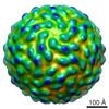 5117MC M: map data used to model this data C: citing same article ( |
|---|---|
| Similar structure data |
-Links
 PDBj
PDBj


- Assembly
| Deposited unit | 
|
|---|---|
| 1 | x 60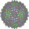
|
| 2 |
|
| 3 | x 5
|
| 4 | x 6
|
| 5 | 
|
| Symmetry | Point symmetry: (Schoenflies symbol: I (icosahedral)) |
-Components
| #1: Protein | Mass: 43904.410 Da / Num. of mol.: 3 Source method: isolated from a genetically manipulated source Source: (gene. exp.) Dengue virus 2 / Cell line (production host): C6/36 mosquito cells / Production host:  #2: Protein | Mass: 9261.531 Da / Num. of mol.: 3 Source method: isolated from a genetically manipulated source Source: (gene. exp.) Dengue virus 2 / Cell line (production host): C6/36 mosquito cells / Production host: |
|---|
-Experimental details
-Experiment
| Experiment | Method: ELECTRON MICROSCOPY |
|---|---|
| EM experiment | Aggregation state: PARTICLE / 3D reconstruction method: single particle reconstruction |
- Sample preparation
| Component |
| |||||||||||||||
|---|---|---|---|---|---|---|---|---|---|---|---|---|---|---|---|---|
| Details of virus | Empty: NO / Enveloped: YES / Host category: VERTEBRATES / Isolate: STRAIN / Type: VIRION | |||||||||||||||
| Virus shell | Triangulation number (T number): 3 | |||||||||||||||
| Buffer solution | pH: 6 / Details: 6 mM Tris-HCL 25 mM MES 120 mM NaCl 1 mM EDTA | |||||||||||||||
| Specimen | Embedding applied: NO / Shadowing applied: NO / Staining applied: NO / Vitrification applied: YES / Details: 6 mM Tris-HCL 25 mM MES 120 mM NaCl 1 mM EDTA | |||||||||||||||
| Vitrification | Cryogen name: ETHANE Method: A small vial of ethane is placed inside a larger liquid nitrogen reservoir. The grid holding a few microliters of the sample is held in place at the bottom of a plunger by the means of fine ...Method: A small vial of ethane is placed inside a larger liquid nitrogen reservoir. The grid holding a few microliters of the sample is held in place at the bottom of a plunger by the means of fine tweezers. Once the ethane in the vial is completely frozen, it needs to be slightly melted. When the liquid ethane is ready, a piece of filter paper is then pressed against the sample to blot of excess buffer, sufficient to leave a thin layer on the grid. After a predetermined time, the filter paper is removed, and the plunger is allowed to drop into the liquid ethane. Once the grid enters the liquid ethane, the sample is rapidly frozen, and the grid is transferred under liquid nitrogen to a storage box immersed liquid nitrogen for later use in the microscope. |
- Electron microscopy imaging
| Microscopy | Model: FEI/PHILIPS CM200FEG / Date: Jun 13, 2008 |
|---|---|
| Electron gun | Electron source:  FIELD EMISSION GUN / Accelerating voltage: 200 kV / Illumination mode: FLOOD BEAM FIELD EMISSION GUN / Accelerating voltage: 200 kV / Illumination mode: FLOOD BEAM |
| Electron lens | Mode: BRIGHT FIELD / Nominal magnification: 50000 X / Nominal defocus max: 3800 nm / Nominal defocus min: 1800 nm / Camera length: 0 mm |
| Specimen holder | Specimen holder model: GATAN LIQUID NITROGEN / Specimen holder type: Eucentric / Tilt angle max: 0 ° / Tilt angle min: 0 ° |
| Image recording | Electron dose: 25 e/Å2 / Film or detector model: GATAN MULTISCAN |
| Image scans | Num. digital images: 45 |
| Radiation | Protocol: SINGLE WAVELENGTH / Monochromatic (M) / Laue (L): M / Scattering type: x-ray |
| Radiation wavelength | Relative weight: 1 |
- Processing
| EM software |
| ||||||||||||
|---|---|---|---|---|---|---|---|---|---|---|---|---|---|
| CTF correction | Details: Each particle | ||||||||||||
| Symmetry | Point symmetry: I (icosahedral) | ||||||||||||
| 3D reconstruction | Resolution: 22 Å / Resolution method: FSC 0.5 CUT-OFF / Num. of particles: 635 / Symmetry type: POINT | ||||||||||||
| Atomic model building | Space: REAL / Target criteria: density match | ||||||||||||
| Atomic model building | PDB-ID: 3C5X Accession code: 3C5X / Source name: PDB / Type: experimental model | ||||||||||||
| Refinement step | Cycle: LAST
|