 Movie
Movie Controller

[English] 日本語
 Yorodumi
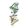
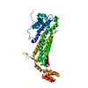
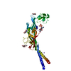
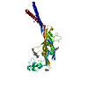
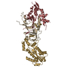
- PDB-2vaz: Model of the S15-mRNA complex fitted into the cryo-EM map of the ... -
+ Open data

- Basic information
| Entry | Database: PDB / ID: 2vaz | ||||||
|---|---|---|---|---|---|---|---|
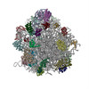
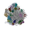
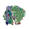
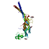
| Title | Model of the S15-mRNA complex fitted into the cryo-EM map of the 70S entrapment complex. | ||||||
 Components Components |
| ||||||
 Keywords Keywords | TRANSLATION / PLATFORM-BINDING CENTER / GENE EXPRESSION REGULATION / RIBOSOMAL PROTEIN / RIBONUCLEOPROTEIN / PROTEIN SYNTHESIS / TRANSLATION INITIATION / RIBOSOME / RIBOSWITCH / RNA-BINDING / RRNA-BINDING / RNA PSEUDOKNOT / MRNA STRUCTURE / REPRESSOR PROTEIN | ||||||
| Function / homology |  Function and homology information Function and homology informationregulation of translation / ribosomal small subunit assembly / small ribosomal subunit rRNA binding / cytosolic small ribosomal subunit / cytoplasmic translation / rRNA binding / structural constituent of ribosome / translation / cytoplasm / cytosol Similarity search - Function | ||||||
| Biological species |  | ||||||
| Method | ELECTRON MICROSCOPY / single particle reconstruction / cryo EM / Resolution: 10 Å | ||||||
| Model type details | P ATOMS ONLY, CHAIN A ; CA ATOMS ONLY, CHAIN F | ||||||
 Authors Authors | Marzi, S. / Myasnikov, A.G. / Serganov, A. / Ehresmann, C. / Romby, P. / Yusupov, M. / Klaholz, B.P. | ||||||
 Citation Citation | Journal: Cell / Year: 2007 Title: Structured mRNAs regulate translation initiation by binding to the platform of the ribosome. Authors: Stefano Marzi / Alexander G Myasnikov / Alexander Serganov / Chantal Ehresmann / Pascale Romby / Marat Yusupov / Bruno P Klaholz /  Abstract: Gene expression can be regulated at the level of initiation of protein biosynthesis via structural elements present at the 5' untranslated region of mRNAs. These folded mRNA segments may bind to the ...Gene expression can be regulated at the level of initiation of protein biosynthesis via structural elements present at the 5' untranslated region of mRNAs. These folded mRNA segments may bind to the ribosome, thus blocking translation until the mRNA unfolds. Here, we report a series of cryo-electron microscopy snapshots of ribosomal complexes directly visualizing either the mRNA structure blocked by repressor protein S15 or the unfolded, active mRNA. In the stalled state, the folded mRNA prevents the start codon from reaching the peptidyl-tRNA (P) site inside the ribosome. Upon repressor release, the mRNA unfolds and moves into the mRNA channel allowing translation initiation. A comparative structure and sequence analysis suggests the existence of a universal stand-by site on the ribosome (the 30S platform) dedicated for binding regulatory 5' mRNA elements. Different types of mRNA structures may be accommodated during translation preinitiation and regulate gene expression by transiently stalling the ribosome. | ||||||
| History |
|
- Structure visualization
| Movie |
Movie viewer |
|---|---|
| Structure viewer | Molecule: MolmilJmol/JSmol |
- Downloads & links
-Download
| PDBx/mmCIF format | 2vaz.cif.gz | 21.6 KB | Display | PDBx/mmCIF format |
|---|---|---|---|---|
| PDB format | pdb2vaz.ent.gz | 7.5 KB | Display | PDB format |
| PDBx/mmJSON format | 2vaz.json.gz | Tree view | PDBx/mmJSON format | |
| Others |  Other downloads Other downloads |
-Validation report
| Summary document | 2vaz_validation.pdf.gz | 725.6 KB | Display | wwPDB validaton report |
|---|---|---|---|---|
| Full document | 2vaz_full_validation.pdf.gz | 725.1 KB | Display | |
| Data in XML | 2vaz_validation.xml.gz | 8.3 KB | Display | |
| Data in CIF | 2vaz_validation.cif.gz | 11 KB | Display | |
| Arichive directory | https://data.pdbj.org/pub/pdb/validation_reports/va/2vazftp://data.pdbj.org/pub/pdb/validation_reports/va/2vaz | HTTPS FTP |
-Related structure data
| Related structure data | 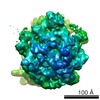 1391MC M: map data used to model this data C: citing same article ( |
|---|---|
| Similar structure data |
-Links
 PDBj
PDBj


- Assembly
| Deposited unit | 
|
|---|---|
| 1 |
|
-Components
| #1: RNA chain | Mass: 59427.113 Da / Num. of mol.: 1 / Fragment: HAIRPIN LOOP DOMAIN I AND PSEUDOKNOT / Source method: obtained synthetically / Source: (synth.) |
|---|---|
| #2: Protein | Mass: 10290.816 Da / Num. of mol.: 1 Source method: isolated from a genetically manipulated source Source: (gene. exp.) |
-Experimental details
-Experiment
| Experiment | Method: ELECTRON MICROSCOPY |
|---|---|
| EM experiment | Aggregation state: PARTICLE / 3D reconstruction method: single particle reconstruction |
- Sample preparation
| Component | Name: 70S PRE-INITIATION COMPLEX ENTRAPPED BY S15- RPSOMRNA / Type: RIBOSOME |
|---|---|
| Buffer solution | Name: 20 MM TRIS-HCL PH 7.5, 60 MM KCL, 1MM DTT, 7.5 MM MGCL2 pH: 7.5 Details: 20 MM TRIS-HCL PH 7.5, 60 MM KCL, 1MM DTT, 7.5 MM MGCL2 |
| Specimen | Conc.: 0.5 mg/ml / Embedding applied: NO / Shadowing applied: NO / Staining applied: NO / Vitrification applied: YES |
| Specimen support | Details: HOLEY CARBON |
| Vitrification | Instrument: HOMEMADE PLUNGER / Cryogen name: ETHANE Details: BLOT FOR 2 SECONDS BEFORE PLUNGING IN LIQUID ETHANE USING A HOME-MADE CRYO- PLUNGER |
- Electron microscopy imaging
| Experimental equipment |  Model: Tecnai F20 / Image courtesy: FEI Company |
|---|---|
| Microscopy | Model: FEI TECNAI F20 / Date: Jan 5, 2005 |
| Electron gun | Electron source:  FIELD EMISSION GUN / Accelerating voltage: 200 kV / Illumination mode: FLOOD BEAM FIELD EMISSION GUN / Accelerating voltage: 200 kV / Illumination mode: FLOOD BEAM |
| Electron lens | Mode: BRIGHT FIELD / Nominal magnification: 50000 X / Calibrated magnification: 51484 X / Nominal defocus max: 3500 nm / Nominal defocus min: 800 nm / Cs: 2 mm |
| Specimen holder | Temperature: 77 K |
| Image recording | Electron dose: 20 e/Å2 / Film or detector model: KODAK SO-163 FILM |
| Image scans | Num. digital images: 67 |
| Radiation wavelength | Relative weight: 1 |
- Processing
| EM software |
| |||||||||||||||||||||
|---|---|---|---|---|---|---|---|---|---|---|---|---|---|---|---|---|---|---|---|---|---|---|
| CTF correction | Details: INDIVIDUAL PARTICLES | |||||||||||||||||||||
| Symmetry | Point symmetry: C1 (asymmetric) | |||||||||||||||||||||
| 3D reconstruction | Method: MANUAL FITTING / Resolution: 10 Å / Num. of particles: 31415 / Nominal pixel size: 3 Å / Actual pixel size: 3 Å Details: THE COORDINATES IN THIS ENTRY WERE GENERATED FROM ELECTRON MICROSCOPY DATA. PROTEIN DATA BANK CONVENTIONS REQUIRE THAT CRYST1 AND SCALE RECORDS BE INCLUDED, BUT THE VALUES ON THESE RECORDS ...Details: THE COORDINATES IN THIS ENTRY WERE GENERATED FROM ELECTRON MICROSCOPY DATA. PROTEIN DATA BANK CONVENTIONS REQUIRE THAT CRYST1 AND SCALE RECORDS BE INCLUDED, BUT THE VALUES ON THESE RECORDS ARE MEANINGLESS. THE MRNA HAS BEEN MODELED USING MANIP (MASSIRE AND WESTHOF). Symmetry type: POINT | |||||||||||||||||||||
| Atomic model building | Protocol: OTHER / Space: REAL Details: METHOD--MANUAL REFINEMENT PROTOCOL--X-RAY AND HOMOLOGY MODELS | |||||||||||||||||||||
| Atomic model building |
| |||||||||||||||||||||
| Refinement | Highest resolution: 10 Å | |||||||||||||||||||||
| Refinement step | Cycle: LAST / Highest resolution: 10 Å
|

