 Movie
Movie Controller

[English] 日本語
 Yorodumi
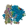
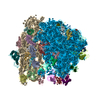
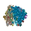
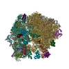
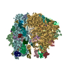
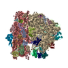
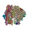
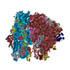
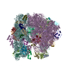
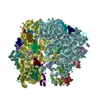
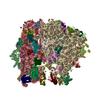
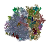
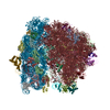
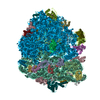
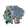
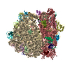
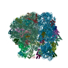
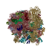
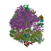
- PDB-4v4q: Crystal structure of the bacterial ribosome from Escherichia coli... -
+ Open data

- Basic information
| Entry | Database: PDB / ID: 4v4q | |||||||||
|---|---|---|---|---|---|---|---|---|---|---|
| Title | Crystal structure of the bacterial ribosome from Escherichia coli at 3.5 A resolution. | |||||||||
 Components Components |
| |||||||||
 Keywords Keywords |  RIBOSOME / RNA-protein complex / 30S RIBOSOMAL SUBUNIT RIBOSOME / RNA-protein complex / 30S RIBOSOMAL SUBUNIT | |||||||||
| Function / homology |  Function and homology information Function and homology informationnegative regulation of cytoplasmic translational initiation / stringent response / mRNA base-pairing translational repressor activity / ornithine decarboxylase inhibitor activity / misfolded RNA binding / transcription antitermination factor activity, RNA binding / Group I intron splicing / RNA folding / transcriptional attenuation / endoribonuclease inhibitor activity ...negative regulation of cytoplasmic translational initiation / stringent response / mRNA base-pairing translational repressor activity / ornithine decarboxylase inhibitor activity / misfolded RNA binding / transcription antitermination factor activity, RNA binding / Group I intron splicing / RNA folding / transcriptional attenuation / endoribonuclease inhibitor activity / RNA-binding transcription regulator activity / positive regulation of ribosome biogenesis / negative regulation of cytoplasmic translation / translational termination / DnaA-L2 complex / four-way junction DNA binding / translation repressor activity / negative regulation of translational initiation / translational initiation / negative regulation of DNA-templated DNA replication initiation / regulation of mRNA stability / ribosome assembly / mRNA regulatory element binding translation repressor activity / response to reactive oxygen species / assembly of large subunit precursor of preribosome / transcription elongation factor complex / positive regulation of RNA splicing / DNA endonuclease activity / : / cytosolic ribosome assembly / regulation of DNA-templated transcription elongation / transcription antitermination / regulation of cell growth / maintenance of translational fidelity / DNA-templated transcription termination / response to radiation / mRNA 5'-UTR binding / ribosomal small subunit biogenesis / small ribosomal subunit rRNA binding / ribosomal small subunit assembly / ribosomal large subunit assembly / cytosolic small ribosomal subunit / large ribosomal subunit rRNA binding / ribosome binding / large ribosomal subunit / ribosome biogenesis / regulation of translation / small ribosomal subunit / 5S rRNA binding / cytoplasmic translation / cytosolic large ribosomal subunit / transferase activity / negative regulation of translation / tRNA binding / molecular adaptor activity / rRNA binding / ribosome / structural constituent of ribosome / translation / response to antibiotic / mRNA binding / negative regulation of DNA-templated transcription / DNA binding / RNA binding / zinc ion binding / membrane / cytosol / cytoplasmSimilarity search - Function | |||||||||
| Biological species |  Escherichia coli (E. coli) Escherichia coli (E. coli) | |||||||||
| Method | X-RAY DIFFRACTION / SYNCHROTRON / MOLECULAR REPLACEMENT / Resolution: 3.46 Å | |||||||||
 Authors Authors | Schuwirth, B.S. / Borovinskaya, M.A. / Hau, C.W. / Zhang, W. / Vila-Sanjurjo, A. / Holton, J.M. / Cate, J.H.D. | |||||||||
 Citation Citation | Journal: Science / Year: 2005 Title: Structures of the bacterial ribosome at 3.5 A resolution. Authors: Barbara S Schuwirth / Maria A Borovinskaya / Cathy W Hau / Wen Zhang / Antón Vila-Sanjurjo / James M Holton / Jamie H Doudna Cate /  Abstract: We describe two structures of the intact bacterial ribosome from Escherichia coli determined to a resolution of 3.5 angstroms by x-ray crystallography. These structures provide a detailed view of the ...We describe two structures of the intact bacterial ribosome from Escherichia coli determined to a resolution of 3.5 angstroms by x-ray crystallography. These structures provide a detailed view of the interface between the small and large ribosomal subunits and the conformation of the peptidyl transferase center in the context of the intact ribosome. Differences between the two ribosomes reveal a high degree of flexibility between the head and the rest of the small subunit. Swiveling of the head of the small subunit observed in the present structures, coupled to the ratchet-like motion of the two subunits observed previously, suggests a mechanism for the final movements of messenger RNA (mRNA) and transfer RNAs (tRNAs) during translocation. | |||||||||
| History |
| |||||||||
| Remark 300 | BIOMOLECULE: 1, 2 THIS ENTRY CONTAINS PART OF THE CRYSTALLOGRAPHIC ASYMMETRIC UNIT. THE BIOLOGICAL ...BIOMOLECULE: 1, 2 THIS ENTRY CONTAINS PART OF THE CRYSTALLOGRAPHIC ASYMMETRIC UNIT. THE BIOLOGICAL UNIT CONSISTS OF TWO SUBUNITS. THERE ARE 2 BIOLOGICAL UNITS IN THE ASYMMETRIC UNIT. | |||||||||
| Remark 400 | COMPOUND THIS FILE, 2AVY, CONTAINS THE 30S SUBUNIT OF ONE 70S RIBOSOME. THE ENTIRE CRYSTAL ...COMPOUND THIS FILE, 2AVY, CONTAINS THE 30S SUBUNIT OF ONE 70S RIBOSOME. THE ENTIRE CRYSTAL STRUCTURE CONTAINS TWO 70S RIBOSOMES AND ARE DEPOSITED UNDER: 70S RIBOSOME ONE: 2AVY (30S SUBUNIT), 2AW4 (50S SUBUNIT) 70S RIBOSOME TWO: 2AW7 (30S SUBUNIT), 2AWB (50S SUBUNIT) | |||||||||
| Remark 600 | HETEROGEN RESIDUE MO4 at 38 HAS SQUARE-PLANAR HYDRATION OF MG2+. RESIDUES MO4 at ...HETEROGEN RESIDUE MO4 at 38 HAS SQUARE-PLANAR HYDRATION OF MG2+. RESIDUES MO4 at 10,16,30,31,34,40,43,44,51 HAVE 2 VICINAL WATERS MISSING FROM HYDRATION OF MG2+. RESIDUES MO3 at 11,48 HAVE ALL WATER BONDS MUTUALLY ORTHOGONAL TO OTHER MG2+-WATER BONDS. RESIDUES MO3 at 4,9,53 HAVE ALL WATERS AND MG2+ IN-PLANE. RESIDUE MO2 at 35 HAS ALL WATERS AND MG2+ ORTHOGONAL. |
- Structure visualization
| Structure viewer | Molecule: MolmilJmol/JSmol |
|---|
- Downloads & links
-Download
| PDBx/mmCIF format | 4v4q.cif.gz | 6.4 MB | Display | PDBx/mmCIF format |
|---|---|---|---|---|
| PDB format | pdb4v4q.ent.gz | Display | PDB format | |
| PDBx/mmJSON format | 4v4q.json.gz | Tree view | PDBx/mmJSON format | |
| Others |  Other downloads Other downloads |
-Validation report
| Arichive directory | https://data.pdbj.org/pub/pdb/validation_reports/v4/4v4qftp://data.pdbj.org/pub/pdb/validation_reports/v4/4v4q | HTTPS FTP |
|---|
-Related structure data
| Related structure data |  1pns 1pnu S: Starting model for refinement |
|---|---|
| Similar structure data |

-Links
 PDBj
PDBj


- Assembly
| Deposited unit | 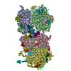
| ||||||||
|---|---|---|---|---|---|---|---|---|---|
| 1 | 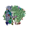
| ||||||||
| 2 | 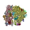
| ||||||||
| Unit cell |
|
-Components
-RNA chain , 3 types, 6 molecules AACABADABBDB
| #1: RNA chain | Mass: 499690.031 Da / Num. of mol.: 2 / Source method: isolated from a natural source / Source: (natural) Escherichia coli (E. coli) / Strain: MRE600 / References: GenBank: 33357879#22: RNA chain | Mass: 38790.090 Da / Num. of mol.: 2 / Source method: isolated from a natural source / Source: (natural) Escherichia coli (E. coli) / Strain: MRE600 / References: GenBank: 33357928#23: RNA chain | Mass: 941612.375 Da / Num. of mol.: 2 / Source method: isolated from a natural source / Source: (natural) Escherichia coli (E. coli) / Strain: MRE600 / References: GenBank: 33357927 |
|---|
-30S ribosomal protein ... , 20 types, 40 molecules ACCCADCDAECEAFCFAGCGAHCHAICIAJCJAKCKALCLAMCMANCNAOCOAPCPAQCQ...
| #2: Protein | Mass: 25900.117 Da / Num. of mol.: 2 / Source method: isolated from a natural source / Source: (natural) Escherichia coli (E. coli) / Strain: MRE600 / References: UniProt: P0A7V3#3: Protein | Mass: 23383.002 Da / Num. of mol.: 2 / Source method: isolated from a natural source / Source: (natural) Escherichia coli (E. coli) / Strain: MRE600 / References: UniProt: P0A7V8#4: Protein | Mass: 17498.203 Da / Num. of mol.: 2 / Source method: isolated from a natural source / Source: (natural) Escherichia coli (E. coli) / Strain: MRE600 / References: UniProt: P0A7W1#5: Protein | Mass: 15727.512 Da / Num. of mol.: 2 / Source method: isolated from a natural source / Source: (natural) Escherichia coli (E. coli) / Strain: MRE600 / References: UniProt: P02358#6: Protein | Mass: 19923.959 Da / Num. of mol.: 2 / Source method: isolated from a natural source / Source: (natural) Escherichia coli (E. coli) / Strain: MRE600 / References: UniProt: P02359#7: Protein | Mass: 14015.361 Da / Num. of mol.: 2 / Source method: isolated from a natural source / Source: (natural) Escherichia coli (E. coli) / Strain: MRE600 / References: UniProt: P0A7W7#8: Protein | Mass: 14755.074 Da / Num. of mol.: 2 / Source method: isolated from a natural source / Source: (natural) Escherichia coli (E. coli) / Strain: MRE600 / References: UniProt: P0A7X3#9: Protein | Mass: 11755.597 Da / Num. of mol.: 2 / Source method: isolated from a natural source / Source: (natural) Escherichia coli (E. coli) / Strain: MRE600 / References: UniProt: P0A7R5#10: Protein | Mass: 13739.778 Da / Num. of mol.: 2 / Source method: isolated from a natural source / Source: (natural) Escherichia coli (E. coli) / Strain: MRE600 / References: UniProt: P0A7R9#11: Protein | Mass: 13636.961 Da / Num. of mol.: 2 / Source method: isolated from a natural source / Source: (natural) Escherichia coli (E. coli) / Strain: MRE600 / References: UniProt: P0A7S3#12: Protein | Mass: 12997.271 Da / Num. of mol.: 2 / Source method: isolated from a natural source / Source: (natural) Escherichia coli (E. coli) / Strain: MRE600 / References: UniProt: P0A7S9#13: Protein | Mass: 11475.364 Da / Num. of mol.: 2 / Source method: isolated from a natural source / Source: (natural) Escherichia coli (E. coli) / Strain: MRE600 / References: UniProt: P02370, UniProt: P0AG59*PLUS#14: Protein | Mass: 10319.882 Da / Num. of mol.: 2 / Source method: isolated from a natural source / Source: (natural) Escherichia coli (E. coli) / Strain: MRE600 / References: UniProt: P02371, UniProt: P0ADZ4*PLUS#15: Protein | Mass: 9207.572 Da / Num. of mol.: 2 / Source method: isolated from a natural source / Source: (natural) Escherichia coli (E. coli) / Strain: MRE600 / References: UniProt: P0A7T3#16: Protein | Mass: 9593.296 Da / Num. of mol.: 2 / Source method: isolated from a natural source / Source: (natural) Escherichia coli (E. coli) / Strain: MRE600 / References: UniProt: P02373, UniProt: P0AG63*PLUS#17: Protein | Mass: 8874.276 Da / Num. of mol.: 2 / Source method: isolated from a natural source / Source: (natural) Escherichia coli (E. coli) / Strain: MRE600 / References: UniProt: P0A7T7#18: Protein | Mass: 10324.160 Da / Num. of mol.: 2 / Source method: isolated from a natural source / Source: (natural) Escherichia coli (E. coli) / Strain: MRE600 / References: UniProt: P0A7U3#19: Protein | Mass: 9577.268 Da / Num. of mol.: 2 / Source method: isolated from a natural source / Source: (natural) Escherichia coli (E. coli) / Strain: MRE600 / References: UniProt: P0A7U7#20: Protein | Mass: 26650.475 Da / Num. of mol.: 2 / Source method: isolated from a natural source / Source: (natural) Escherichia coli (E. coli) / Strain: MRE600 / References: UniProt: P0A7V0#21: Protein | Mass: 8524.039 Da / Num. of mol.: 2 / Source method: isolated from a natural source / Source: (natural) Escherichia coli (E. coli) / Strain: MRE600 / References: UniProt: P68679 |
|---|
+50S ribosomal protein ... , 29 types, 58 molecules BVDVBCDCBDDDBEDEBFDFBGDGBHDHBJDJBKDKBLDLBMDMBNDNBODOBPDPBQDQ...
-Non-polymers , 2 types, 1970 molecules 

| #53: Chemical | ChemComp-MG /  Mass: 24.305 Da / Num. of mol.: 343 / Source method: obtained synthetically / Formula: Mg Mass: 24.305 Da / Num. of mol.: 343 / Source method: obtained synthetically / Formula: Mg#54: Water | ChemComp-HOH / | WaterMass: 18.015 Da / Num. of mol.: 1627 / Source method: isolated from a natural source / Formula: H2O |
|---|
-Experimental details
-Experiment
| Experiment | Method: X-RAY DIFFRACTION / Number of used crystals: 17 |
|---|
- Sample preparation
| Crystal | Density Matthews: 3.41 Å3/Da / Density % sol: 66.85 % | ||||||||||||||||||||||||||||||||||||||||||||||||||||||||
|---|---|---|---|---|---|---|---|---|---|---|---|---|---|---|---|---|---|---|---|---|---|---|---|---|---|---|---|---|---|---|---|---|---|---|---|---|---|---|---|---|---|---|---|---|---|---|---|---|---|---|---|---|---|---|---|---|---|
| Crystal grow | Temperature: 277 K / Method: batch / pH: 7.5 Details: MPD, PEG 8000, MgCl2, NH4Cl, spermine, spermidine, TRIS, EDTA, pH 7.5, batch, temperature 277K | ||||||||||||||||||||||||||||||||||||||||||||||||||||||||
| Components of the solutions |
|
-Data collection
| Diffraction | Mean temperature: 100 K |
|---|---|
| Diffraction source | Source: SYNCHROTRON / Site: ALS / Beamline: 12.3.1 |
| Detector | Type: ADSC QUANTUM 315 / Detector: CCD / Date: Jan 12, 2004 |
| Radiation | Protocol: SINGLE WAVELENGTH / Monochromatic (M) / Laue (L): M / Scattering type: x-ray |
| Radiation wavelength | Relative weight: 1 |
| Reflection | Resolution: 3.46→70 Å / Num. obs: 693093 / % possible obs: 92.3 % / Observed criterion σ(I): -3 / Redundancy: 6.2 % / Biso Wilson estimate: 58.1 Å2 / Rmerge(I) obs: 0.145 / Net I/σ(I): 7.4 |
| Reflection shell | Resolution: 3.46→3.55 Å / Rmerge(I) obs: 0.594 / Mean I/σ(I) obs: 2 / Num. unique obs: 49172 / % possible all: 87.9 |
- Processing
| Software |
| |||||||||||||||||||||||||
|---|---|---|---|---|---|---|---|---|---|---|---|---|---|---|---|---|---|---|---|---|---|---|---|---|---|---|
| Refinement | Method to determine structure: MOLECULAR REPLACEMENT Starting model: pdb entries 1PNS, 1PNU Resolution: 3.46→70 Å / Isotropic thermal model: Grouped by residue / σ(F): 0 / Stereochemistry target values: torsional dynamics
| |||||||||||||||||||||||||
| Displacement parameters | Biso mean: 58.688 Å2 | |||||||||||||||||||||||||
| Refinement step | Cycle: LAST / Resolution: 3.46→70 Å
| |||||||||||||||||||||||||
| Refine LS restraints |
| |||||||||||||||||||||||||
| LS refinement shell |
|
