 Movie
Movie Controller

[English] 日本語
 Yorodumi
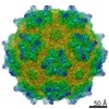
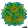
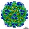
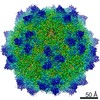
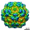
- EMDB-22412: Adeno-associated virus strain AAV7 capsid icosahedral structure -
+ Open data

- Basic information
| Entry | Database: EMDB / ID: EMD-22412 | |||||||||
|---|---|---|---|---|---|---|---|---|---|---|
| Title | Adeno-associated virus strain AAV7 capsid icosahedral structure | |||||||||
 Map data Map data | ||||||||||
 Sample Sample |
| |||||||||
 Keywords Keywords | AAV /  parvovirus / Parvoviridae / adeno-associated virus / capsid / VIRUS parvovirus / Parvoviridae / adeno-associated virus / capsid / VIRUS | |||||||||
| Function / homology | Phospholipase A2-like domain / Phospholipase A2-like domain / Parvovirus coat protein VP2 / Parvovirus coat protein VP1/VP2 / Parvovirus coat protein VP2 / Capsid/spike protein, ssDNA virus / T=1 icosahedral viral capsid / structural molecule activity / Capsid protein Function and homology information Function and homology information | |||||||||
| Biological species |  Adeno-associated virus - 7 Adeno-associated virus - 7 | |||||||||
| Method | single particle reconstruction / cryo EM / Resolution: 2.7 Å | |||||||||
 Authors Authors | Firlar E / Yost SA | |||||||||
 Citation Citation | Journal: Acta Crystallogr F Struct Biol Commun / Year: 2020 Title: Structure of the AAVhu.37 capsid by cryoelectron microscopy. Authors: Jason T Kaelber / Samantha A Yost / Keith A Webber / Emre Firlar / Ye Liu / Olivier Danos / Andrew C Mercer /  Abstract: Adeno-associated viruses (AAVs) are used as in vivo gene-delivery vectors in gene-therapy products and have been heavily investigated for numerous indications. Over 100 naturally occurring AAV ...Adeno-associated viruses (AAVs) are used as in vivo gene-delivery vectors in gene-therapy products and have been heavily investigated for numerous indications. Over 100 naturally occurring AAV serotypes and variants have been isolated from primate samples. Many reports have described unique properties of these variants (for instance, differences in potency, target cell or evasion of the immune response), despite high amino-acid sequence conservation. AAVhu.37 is of interest for clinical applications owing to its proficient transduction of the liver and central nervous system. The sequence identity of the AAVhu.37 VP1 to the well characterized AAVrh.10 serotype, for which no structure is available, is greater than 98%. Here, the structure of the AAVhu.37 capsid at 2.56 Å resolution obtained via single-particle cryo-electron microscopy is presented. | |||||||||
| History |
|
- Structure visualization
| Movie |
Movie viewer |
|---|---|
| Structure viewer | EM map: SurfViewMolmilJmol/JSmol |
| Supplemental images |

- Downloads & links
-EMDB archive
| Map data | emd_22412.map.gz | 106.6 MB | EMDB map data format | |
|---|---|---|---|---|
| Header (meta data) | emd-22412-v30.xmlemd-22412.xml | 20.8 KB 20.8 KB | Display Display | EMDB header |
| FSC (resolution estimation) | emd_22412_fsc.xml | 19 KB | Display | FSC data file |
| Images | 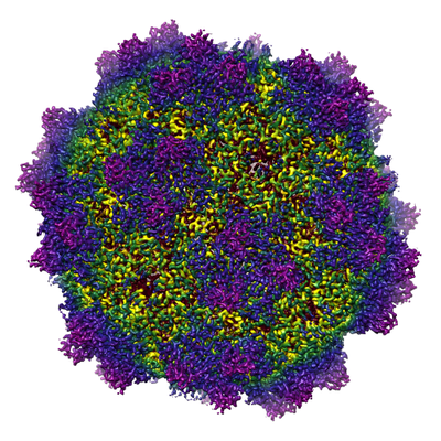 emd_22412.png emd_22412.png | 252.5 KB | ||
| Masks | emd_22412_msk_1.map | 600.7 MB | Mask map | |
| Filedesc metadata | emd-22412.cif.gz | 6.5 KB | ||
| Others | emd_22412_additional_1.map.gzemd_22412_half_map_1.map.gzemd_22412_half_map_2.map.gz | 398.7 MB 483.1 MB 482 MB | ||
| Archive directory |  http://ftp.pdbj.org/pub/emdb/structures/EMD-22412ftp://ftp.pdbj.org/pub/emdb/structures/EMD-22412 http://ftp.pdbj.org/pub/emdb/structures/EMD-22412ftp://ftp.pdbj.org/pub/emdb/structures/EMD-22412 | HTTPS FTP |
-Related structure data
| Related structure data |  7jotMC  6u95C M: atomic model generated by this map C: citing same article ( |
|---|---|
| Similar structure data |


-Links
| EMDB pages | EMDB (EBI/PDBe) / EMDataResource |
|---|---|
| Related items in Molecule of the Month |


-Map
| File | Download / File: emd_22412.map.gz / Format: CCP4 / Size: 600.7 MB / Type: IMAGE STORED AS FLOATING POINT NUMBER (4 BYTES) | ||||||||||||||||||||||||||||||||||||||||||||||||||||||||||||||||||||
|---|---|---|---|---|---|---|---|---|---|---|---|---|---|---|---|---|---|---|---|---|---|---|---|---|---|---|---|---|---|---|---|---|---|---|---|---|---|---|---|---|---|---|---|---|---|---|---|---|---|---|---|---|---|---|---|---|---|---|---|---|---|---|---|---|---|---|---|---|---|
| Voxel size | X=Y=Z: 0.72267 Å | ||||||||||||||||||||||||||||||||||||||||||||||||||||||||||||||||||||
| Density |
| ||||||||||||||||||||||||||||||||||||||||||||||||||||||||||||||||||||
| Symmetry | Space group: 1 | ||||||||||||||||||||||||||||||||||||||||||||||||||||||||||||||||||||
| Details | EMDB XML:
CCP4 map header:
| ||||||||||||||||||||||||||||||||||||||||||||||||||||||||||||||||||||
-Supplemental data
-Mask #1
| File | emd_22412_msk_1.map | ||||||||||||
|---|---|---|---|---|---|---|---|---|---|---|---|---|---|
| Projections & Slices |
| ||||||||||||
| Density Histograms |
 Z
Z Y
Y X
X


-Additional map: #1
| File | emd_22412_additional_1.map | ||||||||||||
|---|---|---|---|---|---|---|---|---|---|---|---|---|---|
| Projections & Slices |
| ||||||||||||
| Density Histograms |


-Half map: #2
| File | emd_22412_half_map_1.map | ||||||||||||
|---|---|---|---|---|---|---|---|---|---|---|---|---|---|
| Projections & Slices |
| ||||||||||||
| Density Histograms |


-Half map: #1
| File | emd_22412_half_map_2.map | ||||||||||||
|---|---|---|---|---|---|---|---|---|---|---|---|---|---|
| Projections & Slices |
| ||||||||||||
| Density Histograms |


- Sample components
-Entire : Adeno-associated virus - 7
| Entire | Name: Adeno-associated virus - 7 |
|---|---|
| Components |
|
-Supramolecule #1: Adeno-associated virus - 7
| Supramolecule | Name: Adeno-associated virus - 7 / type: virus / ID: 1 / Parent: 0 / Macromolecule list: all Details: Virions containing GFP-encoding ssDNA were assembled in transfected HEK293T helper cells. NCBI-ID: 202812 / Sci species name: Adeno-associated virus - 7 / Virus type: VIRION / Virus isolate: SEROTYPE / Virus enveloped: No / Virus empty: No |
|---|---|
| Host (natural) | Organism:  Homo sapiens (human) Homo sapiens (human) |
| Molecular weight | Theoretical: 5 MDa |
| Virus shell | Shell ID: 1 / Diameter: 250.0 Å / T number (triangulation number): 1 |
-Macromolecule #1: Capsid protein
| Macromolecule | Name: Capsid protein / type: protein_or_peptide / ID: 1 / Number of copies: 1 / Enantiomer: LEVO |
|---|---|
| Source (natural) | Organism: Adeno-associated virus - 7 |
| Molecular weight | Theoretical: 58.438547 KDa |
| Recombinant expression | Organism: Homo sapiens (human) |
| Sequence | String: DGVGNASGNW HCDSTWLGDR VITTSTRTWA LPTYNNHLYK QISSETAGST NDNTYFGYST PWGYFDFNRF HCHFSPRDWQ RLINNNWGF RPKKLRFKLF NIQVKEVTTN DGVTTIANNL TSTIQVFSDS EYQLPYVLGS AHQGCLPPFP ADVFMIPQYG Y LTLNNGSQ ...String: DGVGNASGNW HCDSTWLGDR VITTSTRTWA LPTYNNHLYK QISSETAGST NDNTYFGYST PWGYFDFNRF HCHFSPRDWQ RLINNNWGF RPKKLRFKLF NIQVKEVTTN DGVTTIANNL TSTIQVFSDS EYQLPYVLGS AHQGCLPPFP ADVFMIPQYG Y LTLNNGSQ SVGRSSFYCL EYFPSQMLRT GNNFEFSYSF EDVPFHSSYA HSQSLDRLMN PLIDQYLYYL ARTQSNPGGT AG NRELQFY QGGPSTMAEQ AKNWLPGPCF RQQRVSKTLD QNNNSNFAWT GATKYHLNGR NSLVNPGVAM ATHKDDEDRF FPS SGVLIF GKTGATNKTT LENVLMTNEE EIRPTNPVAT EEYGIVSSNL QAANTAAQTQ VVNNQGALPG MVWQNRDVYL QGPI WAKIP HTDGNFHPSP LMGGFGLKHP PPQILIKNTP VPANPPEVFT PAKFASFITQ YSTGQVSVEI EWELQKENSK RWNPE IQYT SNFEKQTGVD FAVDSQGVYS EPRPIGTRYL TRNL UniProtKB: Capsid protein |
-Experimental details
-Structure determination
| Method | cryo EM |
|---|---|
 Processing Processing | single particle reconstruction |
| Aggregation state | particle |
-Sample preparation
| Buffer | pH: 7.4 Component:
Details: PBS | ||||||||||||
|---|---|---|---|---|---|---|---|---|---|---|---|---|---|
| Grid | Model: Quantifoil / Material: COPPER / Support film - Material: CARBON / Support film - topology: CONTINUOUS / Support film - Film thickness: 0.286 / Pretreatment - Type: GLOW DISCHARGE / Pretreatment - Time: 25 sec. / Pretreatment - Atmosphere: AIR / Pretreatment - Pressure: 0.037 kPa | ||||||||||||
| Vitrification | Cryogen name: ETHANE / Chamber humidity: 90 % / Chamber temperature: 298 K / Instrument: LEICA EM GP / Details: Whatman #1 filter paper. | ||||||||||||
| Details | approx. 8*10^14 genome copies per mL |
- Electron microscopy
| Microscope | FEI TALOS ARCTICA |
|---|---|
| Electron beam | Acceleration voltage: 200 kV / Electron source: FIELD EMISSION GUN |
| Electron optics | C2 aperture diameter: 50.0 µm / Calibrated defocus max: 2.836 µm / Calibrated defocus min: 0.244 µm / Illumination mode: FLOOD BEAM / Imaging mode: BRIGHT FIELDBright-field microscopy / Cs: 2.7 mm / Nominal magnification: 130000 |
| Specialist optics | Energy filter - Name: GIF Bioquantum / Energy filter - Slit width: 20 eV |
| Sample stage | Specimen holder model: FEI TITAN KRIOS AUTOGRID HOLDER / Cooling holder cryogen: NITROGEN |
| Image recording | Film or detector model: GATAN K2 SUMMIT (4k x 4k) / Detector mode: COUNTING / Digitization - Dimensions - Width: 3838 pixel / Digitization - Dimensions - Height: 3710 pixel / Digitization - Frames/image: 1-30 / Number grids imaged: 1 / Number real images: 2430 / Average exposure time: 6.0 sec. / Average electron dose: 1.064 e/Å2 |
| Experimental equipment |  Model: Talos Arctica / Image courtesy: FEI Company |
-Image processing
| Particle selection | Number selected: 103227 |
|---|---|
| Startup model | Type of model: NONE |
| Initial angle assignment | Type: PROJECTION MATCHING / Software - Name: RELION (ver. 3.1-beta) |
| Final 3D classification | Software - Name: RELION (ver. 3.1-beta) |
| Final angle assignment | Type: PROJECTION MATCHING / Software - Name: RELION (ver. 3.1-beta) |
| Final reconstruction | Applied symmetry - Point group: I (icosahedral) / Algorithm: FOURIER SPACE / Resolution.type: BY AUTHOR / Resolution: 2.7 Å / Resolution method: FSC 0.143 CUT-OFF / Software - Name: RELION (ver. 3.1-beta) / Number images used: 71811 |
| FSC plot (resolution estimation) |  |
