Journal: J Virol / Year: 2015 Title: Structure of an enteric pathogen, bovine parvovirus. Authors: Shweta Kailasan / Sujata Halder / Brittney Gurda / Heather Bladek / Paul R Chipman / Robert McKenna / Kevin Brown / Mavis Agbandje-McKenna / Abstract: Bovine parvovirus (BPV), the causative agent of respiratory and gastrointestinal disease in cows, is the type member of the Bocaparvovirus genus of the Parvoviridae family. Toward efforts to obtain a ...Bovine parvovirus (BPV), the causative agent of respiratory and gastrointestinal disease in cows, is the type member of the Bocaparvovirus genus of the Parvoviridae family. Toward efforts to obtain a template for the development of vaccines and small-molecule inhibitors for this pathogen, the structure of the BPV capsid, assembled from the major capsid viral protein 2 (VP2), was determined using X-ray crystallography as well as cryo-electron microscopy and three-dimensional image reconstruction (cryo-reconstruction) to 3.2- and 8.8-Å resolutions, respectively. The VP2 region ordered in the crystal structure, from residues 39 to 536, conserves the parvoviral eight-stranded jellyroll motif and an αA helix. The BPV capsid displays common parvovirus features: a channel at and depressions surrounding the 5-fold axes and protrusions surrounding the 3-fold axes. However, rather than a depression centered at the 2-fold axes, a raised surface loop divides this feature in BPV. Additional observed density in the capsid interior in the cryo-reconstructed map, compared to the crystal structure, is interpreted as 10 additional N-terminal residues, residues 29 to 38, that radially extend the channel under the 5-fold axis, as observed for human bocavirus 1 (HBoV1). Surface loops of various lengths and conformations extend from the core jellyroll motif of VP2. These loops confer the unique surface topology of the BPV capsid, making it strikingly different from HBoV1 as well as the type members of other Parvovirinae genera for which structures have been determined. For the type members, regions structurally analogous to those decorating the BPV capsid surface serve as determinants of receptor recognition, tissue and host tropism, pathogenicity, and antigenicity. IMPORTANCE: Bovine parvovirus (BPV), identified in the 1960s in diarrheic calves, is the type member of the Bocaparvovirus genus of the nonenveloped, single-stranded DNA (ssDNA) Parvoviridae family. ...IMPORTANCE: Bovine parvovirus (BPV), identified in the 1960s in diarrheic calves, is the type member of the Bocaparvovirus genus of the nonenveloped, single-stranded DNA (ssDNA) Parvoviridae family. The recent isolation of human bocaparvoviruses from children with severe respiratory and gastrointestinal infections has generated interest in understanding the life cycle and pathogenesis of these emerging viruses. We have determined the high-resolution structure of the BPV capsid assembled from its predominant capsid protein VP2, known to be involved in a myriad of functions during host cell entry, pathogenesis, and antigenicity for other members of the Parvovirinae. Our results show the conservation of the core secondary structural elements and the location of the N-terminal residues for the known bocaparvovirus capsid structures. However, surface loops with high variability in sequence and conformation give BPV a unique capsid surface topology. Similar analogous regions in other Parvovirinae type members are important as determinants of receptor recognition, tissue and host tropism, pathogenicity, and antigenicity.
History
Deposition
Oct 28, 2014
-
Header (metadata) release
Nov 5, 2014
-
Map release
Dec 31, 2014
-
Update
May 27, 2015
-
Current status
May 27, 2015
Processing site: RCSB / Status: Released
-
Structure visualization
Movie
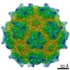
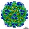
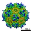
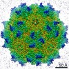
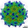
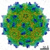
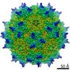
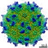
Surface view with section colored by density value
Shell ID: 1 / Name: VP2 / Diameter: 275 Å / T number (triangulation number): 1
-
Experimental details
-
Structure determination
Method
cryo EM
Processing
single particle reconstruction
Aggregation state
particle
-
Sample preparation
Concentration
8.0 mg/mL
Buffer
pH: 7.4 / Details: Tris-HCl
Vitrification
Cryogen name: ETHANE / Chamber humidity: 90 % / Chamber temperature: 120 K / Instrument: FEI VITROBOT MARK IV
-
Electron microscopy
Microscope
FEI TECNAI F20
Alignment procedure
Legacy - Astigmatism: Objective lens astigmatism was corrected at 92,000 times magnification.
Date
Feb 5, 2013
Image recording
Category: CCD / Film or detector model: GATAN ULTRASCAN 4000 (4k x 4k) / Digitization - Sampling interval: 15 µm / Number real images: 63 / Average electron dose: 20 e/Å2
Electron beam
Acceleration voltage: 200 kV / Electron source: FIELD EMISSION GUN
In the structure databanks used in Yorodumi, some data are registered as the other names, "COVID-19 virus" and "2019-nCoV". Here are the details of the virus and the list of structure data.
Jan 31, 2019. EMDB accession codes are about to change! (news from PDBe EMDB page)
EMDB accession codes are about to change! (news from PDBe EMDB page)
The allocation of 4 digits for EMDB accession codes will soon come to an end. Whilst these codes will remain in use, new EMDB accession codes will include an additional digit and will expand incrementally as the available range of codes is exhausted. The current 4-digit format prefixed with “EMD-” (i.e. EMD-XXXX) will advance to a 5-digit format (i.e. EMD-XXXXX), and so on. It is currently estimated that the 4-digit codes will be depleted around Spring 2019, at which point the 5-digit format will come into force.
The EM Navigator/Yorodumi systems omit the EMD- prefix.
Related info.:Q: What is EMD? / ID/Accession-code notation in Yorodumi/EM Navigator
Yorodumi is a browser for structure data from EMDB, PDB, SASBDB, etc.
This page is also the successor to EM Navigator detail page, and also detail information page/front-end page for Omokage search.
The word "yorodu" (or yorozu) is an old Japanese word meaning "ten thousand". "mi" (miru) is to see.
Related info.:EMDB / PDB / SASBDB / Comparison of 3 databanks / Yorodumi Search / Aug 31, 2016. New EM Navigator & Yorodumi / Yorodumi Papers / Jmol/JSmol / Function and homology information / Changes in new EM Navigator and Yorodumi
 Movie
Movie Controller


 Open data
 Basic information
 Map data
 Sample
 Keywords
 Function and homology information
 Bovine parvovirus-1
 Authors
 Citation
 Structure visualization
 Downloads & links
 http://ftp.pdbj.org/pub/emdb/structures/EMD-6168


 Sample components

 Spodoptera frugiperda (fall armyworm) / Recombinant cell: Sf9 / Recombinant plasmid: pFastbac-1
 Processing
 Electron microscopy
 FIELD EMISSION GUN
