 Movie
Movie Controller

[English] 日本語
 Yorodumi
- EMDB-22169: Molecular structure of the core of amyloid-like fibrils formed by... -
+ Open data

- Basic information
| Entry | Database: EMDB / ID: EMD-22169 | |||||||||
|---|---|---|---|---|---|---|---|---|---|---|
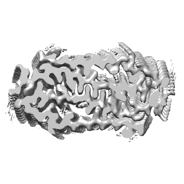
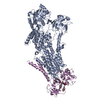
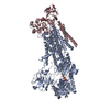
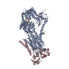
| Title | Molecular structure of the core of amyloid-like fibrils formed by residues 111-214 of FUS | |||||||||
 Map data Map data | ||||||||||
 Sample Sample |
| |||||||||
 Keywords Keywords | Low complexity domain /  Protein aggregation / Amyloid Fibril / RNA BINDING PROTEIN / PROTEIN FIBRIL Protein aggregation / Amyloid Fibril / RNA BINDING PROTEIN / PROTEIN FIBRIL | |||||||||
| Function / homology |  Function and homology information Function and homology informationmRNA stabilization / positive regulation of double-strand break repair via homologous recombination / intracellular non-membrane-bounded organelle / regulation of RNA splicing / Processing of Capped Intron-Containing Pre-mRNA / molecular condensate scaffold activity / mRNA Splicing - Major Pathway / RNA splicing / mRNA 3'-UTR binding / transcription coregulator activity ...mRNA stabilization / positive regulation of double-strand break repair via homologous recombination / intracellular non-membrane-bounded organelle / regulation of RNA splicing / Processing of Capped Intron-Containing Pre-mRNA / molecular condensate scaffold activity / mRNA Splicing - Major Pathway / RNA splicing / mRNA 3'-UTR binding / transcription coregulator activity / protein homooligomerization / amyloid fibril formation / transcription coactivator activity / chromatin binding / regulation of DNA-templated transcription / regulation of transcription by RNA polymerase II / DNA binding / RNA binding / nucleoplasm / identical protein binding / metal ion binding / nucleus / cytoplasmSimilarity search - Function | |||||||||
| Biological species |  Homo sapiens (human) Homo sapiens (human) | |||||||||
| Method | single particle reconstruction / cryo EM / Resolution: 2.62 Å | |||||||||
 Authors Authors | Tycko R / Lee M | |||||||||
 Citation Citation | Journal: Nat Commun / Year: 2020 Title: Molecular structure and interactions within amyloid-like fibrils formed by a low-complexity protein sequence from FUS. Authors: Myungwoon Lee / Ujjayini Ghosh / Kent R Thurber / Masato Kato / Robert Tycko /   Abstract: Protein domains without the usual distribution of amino acids, called low complexity (LC) domains, can be prone to self-assembly into amyloid-like fibrils. Self-assembly of LC domains that are nearly ...Protein domains without the usual distribution of amino acids, called low complexity (LC) domains, can be prone to self-assembly into amyloid-like fibrils. Self-assembly of LC domains that are nearly devoid of hydrophobic residues, such as the 214-residue LC domain of the RNA-binding protein FUS, is particularly intriguing from the biophysical perspective and is biomedically relevant due to its occurrence within neurons in amyotrophic lateral sclerosis, frontotemporal dementia, and other neurodegenerative diseases. We report a high-resolution molecular structural model for fibrils formed by the C-terminal half of the FUS LC domain (FUS-LC-C, residues 111-214), based on a density map with 2.62 Å resolution from cryo-electron microscopy (cryo-EM). In the FUS-LC-C fibril core, residues 112-150 adopt U-shaped conformations and form two subunits with in-register, parallel cross-β structures, arranged with quasi-2 symmetry. All-atom molecular dynamics simulations indicate that the FUS-LC-C fibril core is stabilized by a plethora of hydrogen bonds involving sidechains of Gln, Asn, Ser, and Tyr residues, both along and transverse to the fibril growth direction, including diverse sidechain-to-backbone, sidechain-to-sidechain, and sidechain-to-water interactions. Nuclear magnetic resonance measurements additionally show that portions of disordered residues 151-214 remain highly dynamic in FUS-LC-C fibrils and that fibrils formed by the N-terminal half of the FUS LC domain (FUS-LC-N, residues 2-108) have the same core structure as fibrils formed by the full-length LC domain. These results contribute to our understanding of the molecular structural basis for amyloid formation by FUS and by LC domains in general. | |||||||||
| History |
|
- Structure visualization
| Movie |
Movie viewer |
|---|---|
| Structure viewer | EM map: SurfViewMolmilJmol/JSmol |
| Supplemental images |
- Downloads & links
-EMDB archive
| Map data | emd_22169.map.gz | 11.2 MB | EMDB map data format | |
|---|---|---|---|---|
| Header (meta data) | emd-22169-v30.xmlemd-22169.xml | 13.6 KB 13.6 KB | Display Display | EMDB header |
| Images |  emd_22169.png emd_22169.png | 149.9 KB | ||
| Filedesc metadata | emd-22169.cif.gz | 5.6 KB | ||
| Archive directory |  http://ftp.pdbj.org/pub/emdb/structures/EMD-22169ftp://ftp.pdbj.org/pub/emdb/structures/EMD-22169 http://ftp.pdbj.org/pub/emdb/structures/EMD-22169ftp://ftp.pdbj.org/pub/emdb/structures/EMD-22169 | HTTPS FTP |
-Related structure data
| Related structure data |  6xfmMC M: atomic model generated by this map C: citing same article ( |
|---|---|
| Similar structure data |


-Links
| EMDB pages | EMDB (EBI/PDBe) / EMDataResource |
|---|---|
| Related items in Molecule of the Month |


-Map
| File | Download / File: emd_22169.map.gz / Format: CCP4 / Size: 244.1 MB / Type: IMAGE STORED AS FLOATING POINT NUMBER (4 BYTES) | ||||||||||||||||||||||||||||||||||||||||||||||||||||||||||||||||||||
|---|---|---|---|---|---|---|---|---|---|---|---|---|---|---|---|---|---|---|---|---|---|---|---|---|---|---|---|---|---|---|---|---|---|---|---|---|---|---|---|---|---|---|---|---|---|---|---|---|---|---|---|---|---|---|---|---|---|---|---|---|---|---|---|---|---|---|---|---|---|
| Voxel size | X=Y=Z: 1.074 Å | ||||||||||||||||||||||||||||||||||||||||||||||||||||||||||||||||||||
| Density |
| ||||||||||||||||||||||||||||||||||||||||||||||||||||||||||||||||||||
| Symmetry | Space group: 1 | ||||||||||||||||||||||||||||||||||||||||||||||||||||||||||||||||||||
| Details | EMDB XML:
CCP4 map header:
| ||||||||||||||||||||||||||||||||||||||||||||||||||||||||||||||||||||
-Supplemental data
- Sample components
-Entire : FUS low complexity sequence
| Entire | Name: FUS low complexity sequence |
|---|---|
| Components |
|
-Supramolecule #1: FUS low complexity sequence
| Supramolecule | Name: FUS low complexity sequence / type: complex / ID: 1 / Parent: 0 / Macromolecule list: all Details: C-terminal domain of FUS low complexity domain (111-214) |
|---|---|
| Source (natural) | Organism: Homo sapiens (human) |
| Molecular weight | Theoretical: 40.4 kDa/nm |
-Macromolecule #1: RNA-binding protein FUS
| Macromolecule | Name: RNA-binding protein FUS / type: protein_or_peptide / ID: 1 / Number of copies: 8 / Enantiomer: LEVO |
|---|---|
| Source (natural) | Organism: Homo sapiens (human) |
| Molecular weight | Theoretical: 10.024784 KDa |
| Recombinant expression | Organism:  Escherichia coli BL21(DE3) (bacteria) Escherichia coli BL21(DE3) (bacteria) |
| Sequence | String: GSYGSSSQSS SYGQPQSGSY SQQPSYGGQQ QSYGQQQSYN PPQGYGQQNQ YNSSSGGGGG GGGGGNYGQD QSSMSSGGGS GGGYGNQDQ SGGGGSGGYG QGDRG UniProtKB: RNA-binding protein FUS |
-Experimental details
-Structure determination
| Method | cryo EM |
|---|---|
 Processing Processing | single particle reconstruction |
| Aggregation state | helical array |
-Sample preparation
| Concentration | 0.4 mg/mL |
|---|---|
| Buffer | pH: 7.4 / Component - Concentration: 20.0 mM / Component - Formula: C4H11NO3 / Component - Name: Tris HCl / Details: 20 mM 2-mercaptoethanol, 0.1 mM PMSF |
| Grid | Model: Quantifoil R2/2 / Material: COPPER / Mesh: 300 / Support film - Material: CARBON / Support film - topology: HOLEY / Support film - Film thickness: 12 / Pretreatment - Type: GLOW DISCHARGE / Pretreatment - Time: 60 sec. / Pretreatment - Pressure: 0.039 kPa Details: The grid was glow discharged immediately before use. |
| Vitrification | Cryogen name: ETHANE / Chamber humidity: 99 % / Chamber temperature: 93 K / Instrument: LEICA PLUNGER Details: Preblot for 10 seconds and blot for 5 seconds before plunging. |
- Electron microscopy
| Microscope | FEI TITAN KRIOS |
|---|---|
| Electron beam | Acceleration voltage: 300 kV / Electron source: FIELD EMISSION GUN |
| Electron optics | Illumination mode: FLOOD BEAM / Imaging mode: BRIGHT FIELDBright-field microscopy / Nominal defocus max: 2.5 µm / Nominal defocus min: 1.0 µm / Nominal magnification: 130000 |
| Sample stage | Cooling holder cryogen: NITROGEN |
| Image recording | Film or detector model: GATAN K2 SUMMIT (4k x 4k) / Detector mode: SUPER-RESOLUTION / Digitization - Dimensions - Width: 3800 pixel / Digitization - Dimensions - Height: 3700 pixel / Number grids imaged: 2 / Number real images: 2404 / Average exposure time: 6.0 sec. / Average electron dose: 47.0 e/Å2 Details: 58185 fibril segments were manually selected from 2404 micrographs |
| Experimental equipment |  Model: Titan Krios / Image courtesy: FEI Company |
-Image processing
| Particle selection | Number selected: 499206 Details: 499206 of particles were extracted from the 58185 fibril segments using a 400-pixel box size and 91.6% overlap. |
|---|---|
| Startup model | Type of model: NONE / Details: featureless cylinder |
| Initial angle assignment | Type: ANGULAR RECONSTITUTION / Software - Name: RELION (ver. 3.0) Details: 4.8-Angstrom helical rise and -2.0 degree helical twist |
| Final 3D classification | Number classes: 3 / Software - Name: RELION (ver. 3.0) Details: The major class which contains 69% of the particles was selected for further refinement |
| Final angle assignment | Type: ANGULAR RECONSTITUTION / Software - Name: RELION (ver. 3.0) |
| Final reconstruction | Number classes used: 1 / Applied symmetry - Point group: C1 (asymmetric) / Algorithm: FOURIER SPACE / Resolution.type: BY AUTHOR / Resolution: 2.62 Å / Resolution method: FSC 0.143 CUT-OFF / Software - Name: RELION (ver. 3.0) Details: 3D refinement and post-processing were performed with 21 (screw) symmetry Number images used: 275520 |
| Details | Gatan Imaging Filter (GIF) Quantum LS |
-Atomic model buiding 1
| Details | Manually generated model was fit into the density using PHENIX and UCSF Chimera. Further refinements were performed using Xplor-NIH. |
|---|---|
| Refinement | Protocol: OTHER |
| Output model | PDB-6xfm: |
