 Movie
Movie Controller

[English] 日本語
 Yorodumi
- PDB-7bsq: Cryo-EM structure of a human ATP11C-CDC50A flippase in E1AlF-ADP state -
+ Open data

- Basic information
| Entry | Database: PDB / ID: 7bsq | ||||||||||||
|---|---|---|---|---|---|---|---|---|---|---|---|---|---|
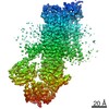
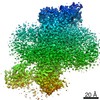
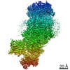
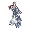
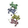
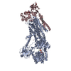
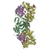
| Title | Cryo-EM structure of a human ATP11C-CDC50A flippase in E1AlF-ADP state | ||||||||||||
 Components Components |
| ||||||||||||
 Keywords Keywords |  MEMBRANE PROTEIN / Flippase / P-type ATPase / P4-ATPase / Phospholipid MEMBRANE PROTEIN / Flippase / P-type ATPase / P4-ATPase / Phospholipid | ||||||||||||
| Function / homology |  Function and homology information Function and homology informationpositive regulation of phospholipid translocation / aminophospholipid flippase activity / aminophospholipid transport / phosphatidylserine flippase activity / protein localization to endosome / phosphatidylserine floppase activity / phospholipid-translocating ATPase complex / ATPase-coupled intramembrane lipid transporter activity / positive regulation of protein exit from endoplasmic reticulum / xenobiotic transmembrane transport ...positive regulation of phospholipid translocation / aminophospholipid flippase activity / aminophospholipid transport / phosphatidylserine flippase activity / protein localization to endosome / phosphatidylserine floppase activity / phospholipid-translocating ATPase complex / ATPase-coupled intramembrane lipid transporter activity / positive regulation of protein exit from endoplasmic reticulum / xenobiotic transmembrane transport / phosphatidylethanolamine flippase activity / P-type phospholipid transporter / phospholipid translocation / azurophil granule membrane / transport vesicle membrane / Ion transport by P-type ATPases / specific granule membrane / monoatomic ion transmembrane transport / positive regulation of neuron projection development / recycling endosome / recycling endosome membrane / late endosome membrane / early endosome membrane / apical plasma membrane / lysosomal membrane / Neutrophil degranulation / endoplasmic reticulum membrane / structural molecule activity / Golgi apparatus / magnesium ion binding / endoplasmic reticulum / ATP hydrolysis activity / ATP binding / membrane / plasma membraneSimilarity search - Function | ||||||||||||
| Biological species |  Homo sapiens (human) Homo sapiens (human) | ||||||||||||
| Method | ELECTRON MICROSCOPY / single particle reconstruction / cryo EM / Resolution: 3.2 Å | ||||||||||||
 Authors Authors | Abe, K. / Nishizawa, T. / Nakanishi, H. | ||||||||||||
| Funding support |  Japan, 3items Japan, 3items
| ||||||||||||
 Citation Citation | Journal: Cell Rep / Year: 2020 Title: Transport Cycle of Plasma Membrane Flippase ATP11C by Cryo-EM. Authors: Hanayo Nakanishi / Tomohiro Nishizawa / Katsumori Segawa / Osamu Nureki / Yoshinori Fujiyoshi / Shigekazu Nagata / Kazuhiro Abe / Abstract: ATP11C, a plasma membrane phospholipid flippase, maintains the asymmetric distribution of phosphatidylserine accumulated in the inner leaflet. Caspase-dependent inactivation of ATP11C is essential ...ATP11C, a plasma membrane phospholipid flippase, maintains the asymmetric distribution of phosphatidylserine accumulated in the inner leaflet. Caspase-dependent inactivation of ATP11C is essential for an apoptotic "eat me" signal, phosphatidylserine exposure, which prompts phagocytes to engulf cells. We show six cryo-EM structures of ATP11C at 3.0-4.0 Å resolution in five different states of the transport cycle. A structural comparison reveals phosphorylation-driven domain movements coupled with phospholipid binding. Three structures of phospholipid-bound states visualize phospholipid translocation accompanied by the rearrangement of transmembrane helices and an unwound portion at the occlusion site, and thus they detail the basis for head group recognition and the locality of the protein-bound acyl chains in transmembrane grooves. Invariant Lys880 and the surrounding hydrogen-bond network serve as a pivot point for helix bending and precise P domain inclination, which is crucial for dephosphorylation. The structures detail key features of phospholipid translocation by ATP11C, and a common basic mechanism for flippases is emerging. | ||||||||||||
| History |
|
- Structure visualization
| Movie |
Movie viewer |
|---|---|
| Structure viewer | Molecule: MolmilJmol/JSmol |
- Downloads & links
-Download
| PDBx/mmCIF format | 7bsq.cif.gz | 259.1 KB | Display | PDBx/mmCIF format |
|---|---|---|---|---|
| PDB format | pdb7bsq.ent.gz | 203.3 KB | Display | PDB format |
| PDBx/mmJSON format | 7bsq.json.gz | Tree view | PDBx/mmJSON format | |
| Others |  Other downloads Other downloads |
-Validation report
| Arichive directory | https://data.pdbj.org/pub/pdb/validation_reports/bs/7bsqftp://data.pdbj.org/pub/pdb/validation_reports/bs/7bsq | HTTPS FTP |
|---|
-Related structure data
| Related structure data | 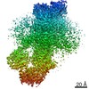 30164MC 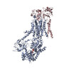 7bspC 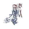 7bssC 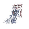 7bsuC 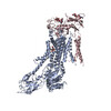 7bsvC 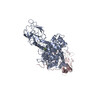 7bswC M: map data used to model this data C: citing same article ( |
|---|---|
| Similar structure data |


-Links
 PDBj
PDBj


- Assembly
| Deposited unit | 
|
|---|---|
| 1 |
|
-Components
-Protein , 2 types, 2 molecules AC
| #1: Protein | Mass: 124403.617 Da / Num. of mol.: 1 / Mutation: delete 7 a.a. at N-term and 38 a.a. at C-term Source method: isolated from a genetically manipulated source Source: (gene. exp.) Homo sapiens (human) / Cell line (production host): Expi293 / Production host: Homo sapiens (human) / References: UniProt: Q8NB49*PLUS |
|---|---|
| #2: Protein | Mass: 40900.801 Da / Num. of mol.: 1 / Mutation: N180Q, S292W Source method: isolated from a genetically manipulated source Source: (gene. exp.) Homo sapiens (human) / Cell line (production host): Expi293 / Production host: Homo sapiens (human) / References: UniProt: Q9NV96*PLUS |
-Sugars , 2 types, 3 molecules 
| #3: Polysaccharide | alpha-D-mannopyranose-(1-3)-2-acetamido-2-deoxy-beta-D-glucopyranose-(1-4)-2-acetamido-2-deoxy-beta- ...alpha-D-mannopyranose-(1-3)-2-acetamido-2-deoxy-beta-D-glucopyranose-(1-4)-2-acetamido-2-deoxy-beta-D-glucopyranose / Mass: 586.542 Da / Num. of mol.: 1 Source method: isolated from a genetically manipulated source |
|---|---|
| #7: Sugar | N-Acetylglucosamine Type: D-saccharide, beta linking / Mass: 221.208 Da / Num. of mol.: 2 Type: D-saccharide, beta linking / Mass: 221.208 Da / Num. of mol.: 2Source method: isolated from a genetically manipulated source Formula: C8H15NO6 |
-Non-polymers , 3 types, 4 molecules 


| #4: Chemical | ChemComp-ALF /  Mass: 102.975 Da / Num. of mol.: 1 / Source method: obtained synthetically / Formula: AlF4 / Feature type: SUBJECT OF INVESTIGATION Mass: 102.975 Da / Num. of mol.: 1 / Source method: obtained synthetically / Formula: AlF4 / Feature type: SUBJECT OF INVESTIGATION |
|---|---|
| #5: Chemical | ChemComp-ADP / Adenosine diphosphate Mass: 427.201 Da / Num. of mol.: 1 / Source method: obtained synthetically / Formula: C10H15N5O10P2 / Feature type: SUBJECT OF INVESTIGATION / Comment: ADP, energy-carrying molecule*YM Mass: 427.201 Da / Num. of mol.: 1 / Source method: obtained synthetically / Formula: C10H15N5O10P2 / Feature type: SUBJECT OF INVESTIGATION / Comment: ADP, energy-carrying molecule*YM |
| #6: Chemical |  Mass: 24.305 Da / Num. of mol.: 2 / Source method: obtained synthetically / Formula: Mg / Feature type: SUBJECT OF INVESTIGATION Mass: 24.305 Da / Num. of mol.: 2 / Source method: obtained synthetically / Formula: Mg / Feature type: SUBJECT OF INVESTIGATION |
-Details
| Has ligand of interest | Y |
|---|---|
| Sequence details | NCBI Reference Sequence accession IDs are NCBI XM_005262405.1 for ATP11C and NP_001340741.2 for CDC50A. |
-Experimental details
-Experiment
| Experiment | Method: ELECTRON MICROSCOPY |
|---|---|
| EM experiment | Aggregation state: PARTICLE / 3D reconstruction method: single particle reconstruction |
- Sample preparation
| Component | Name: human ATP11C-CDC50A flippase / Type: COMPLEX / Entity ID: #1-#2 / Source: RECOMBINANT |
|---|---|
| Molecular weight | Value: 0.18 MDa / Experimental value: NO |
| Source (natural) | Organism: Homo sapiens (human) |
| Source (recombinant) | Organism: Homo sapiens (human) / Cell: Expi293 |
| Buffer solution | pH: 6.5 |
| Specimen | Conc.: 8 mg/ml / Embedding applied: NO / Shadowing applied: NO / Staining applied: NO / Vitrification applied: YES |
| Vitrification | Instrument: FEI VITROBOT MARK IV / Cryogen name: ETHANE / Humidity: 99 % / Chamber temperature: 298 K |
- Electron microscopy imaging
| Experimental equipment |  Model: Titan Krios / Image courtesy: FEI Company |
|---|---|
| Microscopy | Model: TFS KRIOS |
| Electron gun | Electron source: FIELD EMISSION GUN / Accelerating voltage: 300 kV / Illumination mode: FLOOD BEAM |
| Electron lens | Mode: BRIGHT FIELDBright-field microscopy |
| Image recording | Electron dose: 64 e/Å2 / Film or detector model: GATAN K3 (6k x 4k) |
- Processing
| Software |
| ||||||||||||||||||||||||
|---|---|---|---|---|---|---|---|---|---|---|---|---|---|---|---|---|---|---|---|---|---|---|---|---|---|
| EM software |
| ||||||||||||||||||||||||
| CTF correction | Type: PHASE FLIPPING AND AMPLITUDE CORRECTION | ||||||||||||||||||||||||
| 3D reconstruction | Resolution: 3.2 Å / Resolution method: FSC 0.143 CUT-OFF / Num. of particles: 500000 / Symmetry type: POINT | ||||||||||||||||||||||||
| Atomic model building | Protocol: RIGID BODY FIT | ||||||||||||||||||||||||
| Refinement | Cross valid method: NONE Stereochemistry target values: GeoStd + Monomer Library + CDL v1.2 | ||||||||||||||||||||||||
| Displacement parameters | Biso mean: 54.07 Å2 | ||||||||||||||||||||||||
| Refine LS restraints |
|
