 ムービー
ムービー コントローラー


+ データを開く

- 基本情報
基本情報
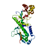
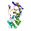
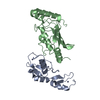
| 登録情報 | データベース: PDB / ID: 6xfm | ||||||
|---|---|---|---|---|---|---|---|
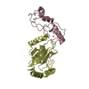
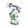
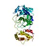
| タイトル | Molecular structure of the core of amyloid-like fibrils formed by residues 111-214 of FUS | ||||||
 要素 要素 | RNA-binding protein FUS FUS FUS | ||||||
 キーワード キーワード | RNA BINDING PROTEIN (RNA結合タンパク質) / PROTEIN FIBRIL / Low complexity domain / Protein aggregation (タンパク質凝集) / Amyloid Fibril (アミロイド) | ||||||
| 機能・相同性 |  機能・相同性情報 機能・相同性情報mRNA stabilization / positive regulation of double-strand break repair via homologous recombination / intracellular non-membrane-bounded organelle / regulation of RNA splicing / Processing of Capped Intron-Containing Pre-mRNA / molecular condensate scaffold activity / mRNA Splicing - Major Pathway / RNA splicing / mRNA 3'-UTR binding / transcription coregulator activity ...mRNA stabilization / positive regulation of double-strand break repair via homologous recombination / intracellular non-membrane-bounded organelle / regulation of RNA splicing / Processing of Capped Intron-Containing Pre-mRNA / molecular condensate scaffold activity / mRNA Splicing - Major Pathway / RNA splicing / mRNA 3'-UTR binding / transcription coregulator activity / protein homooligomerization / amyloid fibril formation / transcription coactivator activity / chromatin binding / regulation of DNA-templated transcription / regulation of transcription by RNA polymerase II / DNA binding / RNA binding / 核質 / identical protein binding / metal ion binding / 細胞核 / 細胞質類似検索 - 分子機能 | ||||||
| 生物種 |  Homo sapiens (ヒト) Homo sapiens (ヒト) | ||||||
| 手法 | 電子顕微鏡法 / 単粒子再構成法 / クライオ電子顕微鏡法 / 解像度: 2.62 Å | ||||||
 データ登録者 データ登録者 | Tycko, R. / Lee, M. / Ghosh, U. / Thurber, K. / Kato, M. | ||||||
 引用 引用 | ジャーナル: Nat Commun / 年: 2020 タイトル: Molecular structure and interactions within amyloid-like fibrils formed by a low-complexity protein sequence from FUS. 著者: Myungwoon Lee / Ujjayini Ghosh / Kent R Thurber / Masato Kato / Robert Tycko /   要旨: Protein domains without the usual distribution of amino acids, called low complexity (LC) domains, can be prone to self-assembly into amyloid-like fibrils. Self-assembly of LC domains that are nearly ...Protein domains without the usual distribution of amino acids, called low complexity (LC) domains, can be prone to self-assembly into amyloid-like fibrils. Self-assembly of LC domains that are nearly devoid of hydrophobic residues, such as the 214-residue LC domain of the RNA-binding protein FUS, is particularly intriguing from the biophysical perspective and is biomedically relevant due to its occurrence within neurons in amyotrophic lateral sclerosis, frontotemporal dementia, and other neurodegenerative diseases. We report a high-resolution molecular structural model for fibrils formed by the C-terminal half of the FUS LC domain (FUS-LC-C, residues 111-214), based on a density map with 2.62 Å resolution from cryo-electron microscopy (cryo-EM). In the FUS-LC-C fibril core, residues 112-150 adopt U-shaped conformations and form two subunits with in-register, parallel cross-β structures, arranged with quasi-2 symmetry. All-atom molecular dynamics simulations indicate that the FUS-LC-C fibril core is stabilized by a plethora of hydrogen bonds involving sidechains of Gln, Asn, Ser, and Tyr residues, both along and transverse to the fibril growth direction, including diverse sidechain-to-backbone, sidechain-to-sidechain, and sidechain-to-water interactions. Nuclear magnetic resonance measurements additionally show that portions of disordered residues 151-214 remain highly dynamic in FUS-LC-C fibrils and that fibrils formed by the N-terminal half of the FUS LC domain (FUS-LC-N, residues 2-108) have the same core structure as fibrils formed by the full-length LC domain. These results contribute to our understanding of the molecular structural basis for amyloid formation by FUS and by LC domains in general. | ||||||
| 履歴 |
|
- 構造の表示
構造の表示
| ムービー |
ムービービューア |
|---|---|
| 構造ビューア | 分子: MolmilJmol/JSmol |
- ダウンロードとリンク
-ダウンロード
| PDBx/mmCIF形式 | 6xfm.cif.gz | 1.1 MB | 表示 | PDBx/mmCIF形式 |
|---|---|---|---|---|
| PDB形式 | pdb6xfm.ent.gz | 947.5 KB | 表示 | PDB形式 |
| PDBx/mmJSON形式 | 6xfm.json.gz | ツリー表示 | PDBx/mmJSON形式 | |
| その他 |  その他のダウンロード その他のダウンロード |
-検証レポート
| アーカイブディレクトリ | https://data.pdbj.org/pub/pdb/validation_reports/xf/6xfmftp://data.pdbj.org/pub/pdb/validation_reports/xf/6xfm | HTTPS FTP |
|---|
-関連構造データ


-リンク
 PDBj
PDBj


- 集合体
集合体
| 登録構造単位 | 
|
|---|---|
| 1 |
|
| モデル数 | 14 |
-要素
| #1: タンパク質 | FUS / 75 kDa DNA-pairing protein / Oncogene FUS / Oncogene TLS / POMp75 / Translocated in liposarcoma protein 分子量: 10024.784 Da / 分子数: 8 / 断片: low complexity domain (UNP residues 111-214) / 由来タイプ: 組換発現 / 由来: (組換発現) Homo sapiens (ヒト) / 遺伝子: FUS, TLS / 発現宿主:  Escherichia coli BL21(DE3) (大腸菌) / 参照: UniProt: P35637 Escherichia coli BL21(DE3) (大腸菌) / 参照: UniProt: P35637 |
|---|
-実験情報
-実験
| 実験 | 手法: 電子顕微鏡法 |
|---|---|
| EM実験 | 試料の集合状態: HELICAL ARRAY / 3次元再構成法: 単粒子再構成法 |
- 試料調製
試料調製
| 構成要素 | 名称: FUS low complexity sequence / タイプ: COMPLEX 詳細: C-terminal domain of FUS low complexity domain (111-214) Entity ID: all / 由来: RECOMBINANT |
|---|---|
| 分子量 | 値: 40.4 kDa/nm / 実験値: YES |
| 由来(天然) | 生物種: Homo sapiens (ヒト) |
| 由来(組換発現) | 生物種: Escherichia coli BL21(DE3) (大腸菌) |
| 緩衝液 | pH: 7.4 / 詳細: 20 mM 2-mercaptoethanol, 0.1 mM PMSF |
| 緩衝液成分 | 濃度: 20 mM / 名称: Tris HCl / 式: C4H11NO3 |
| 試料 | 濃度: 0.4 mg/ml / 包埋: NO / シャドウイング: NO / 染色: NO / 凍結: YES |
| 試料支持 | 詳細: The grid was glow discharged immediately before use. グリッドの材料: COPPER / グリッドのサイズ: 300 divisions/in. / グリッドのタイプ: Quantifoil R2/2 |
| 急速凍結 | 装置: LEICA PLUNGER / 凍結剤: ETHANE / 湿度: 99 % / 凍結前の試料温度: 93 K 詳細: Preblot for 10 seconds and blot for 5 seconds before plunging |
- 電子顕微鏡撮影
| 実験機器 |  モデル: Titan Krios / 画像提供: FEI Company |
|---|---|
| 顕微鏡 | モデル: FEI TITAN KRIOS |
| 電子銃 | 電子線源: FIELD EMISSION GUN / 加速電圧: 300 kV / 照射モード: FLOOD BEAM |
| 電子レンズ | モード: BRIGHT FIELDBright-field microscopy / 倍率(公称値): 130000 X / 最大 デフォーカス(公称値): 2500 nm / 最小 デフォーカス(公称値): 1000 nm |
| 試料ホルダ | 凍結剤: NITROGEN |
| 撮影 | 平均露光時間: 6 sec. / 電子線照射量: 47 e/Å2 / 検出モード: SUPER-RESOLUTION フィルム・検出器のモデル: GATAN K2 SUMMIT (4k x 4k) 撮影したグリッド数: 2 / 実像数: 2404 詳細: 58185 fibril segments were manually selected from 2404 micrographs |
| 画像スキャン | 横: 3800 / 縦: 3700 / 動画フレーム数/画像: 30 |
- 解析
解析
| EMソフトウェア |
| ||||||||||||||||||||||||||||||||||||||||||||||||
|---|---|---|---|---|---|---|---|---|---|---|---|---|---|---|---|---|---|---|---|---|---|---|---|---|---|---|---|---|---|---|---|---|---|---|---|---|---|---|---|---|---|---|---|---|---|---|---|---|---|
| 画像処理 | 詳細: Gatan Imaging Filter (GIF) Quantum LS | ||||||||||||||||||||||||||||||||||||||||||||||||
| CTF補正 | 詳細: Gctf / タイプ: NONE | ||||||||||||||||||||||||||||||||||||||||||||||||
| らせん対称 | 回転角度/サブユニット: -2 ° / 軸方向距離/サブユニット: 4.8 Å / らせん対称軸の対称性: C2 | ||||||||||||||||||||||||||||||||||||||||||||||||
| 粒子像の選択 | 選択した粒子像数: 499206 詳細: 499206 of particles were extracted from the 58185 fibril segments using a 400-pixel box size and 91.6% overlap. | ||||||||||||||||||||||||||||||||||||||||||||||||
| 対称性 | 点対称性: C1 (非対称) | ||||||||||||||||||||||||||||||||||||||||||||||||
| 3次元再構成 | 解像度: 2.62 Å / 解像度の算出法: FSC 0.143 CUT-OFF / 粒子像の数: 275520 / アルゴリズム: FOURIER SPACE 詳細: 3D refinement and post-processing were performed with 21 (screw) symmetry クラス平均像の数: 1 / 対称性のタイプ: POINT | ||||||||||||||||||||||||||||||||||||||||||||||||
| 原子モデル構築 | プロトコル: OTHER 詳細: Manually generated model was fit into the density using PHENIX and UCSF Chimera. Further refinements were performed using Xplor-NIH. |
