 Movie
Movie Controller

[English] 日本語
 Yorodumi
- PDB-6j7c: Crystal structure of proline racemase-like protein from Thermococ... -
+ Open data

- Basic information
| Entry | Database: PDB / ID: 6j7c | ||||||
|---|---|---|---|---|---|---|---|
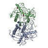
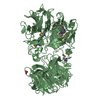
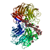
| Title | Crystal structure of proline racemase-like protein from Thermococcus litoralis in complex with proline | ||||||
 Components Components | Proline racemase | ||||||
 Keywords Keywords | ISOMERASE / Proline racemase / Hydroxyproline 2-epimerase / Hyperthermophilic archaea | ||||||
| Function / homology | 4-hydroxyproline epimerase activity / Proline racemase family / Proline racemase / Diaminopimelate Epimerase; Chain A, domain 1 / Diaminopimelate Epimerase; Chain A, domain 1 / Roll / Alpha Beta / PROLINE / Proline racemase Function and homology information Function and homology information | ||||||
| Biological species |   Thermococcus litoralis (archaea) Thermococcus litoralis (archaea) | ||||||
| Method |  X-RAY DIFFRACTION / SYNCHROTRON / MOLECULAR REPLACEMENT / Resolution: 2.7 Å X-RAY DIFFRACTION / SYNCHROTRON / MOLECULAR REPLACEMENT / Resolution: 2.7 Å | ||||||
 Authors Authors | Watanabe, Y. / Watanabe, S. / Itoh, Y. / Watanabe, Y. | ||||||
| Funding support |  Japan, 1items Japan, 1items
| ||||||
 Citation Citation | Journal: Biochem. Biophys. Res. Commun. / Year: 2019 Title: Crystal structure of substrate-bound bifunctional proline racemase/hydroxyproline epimerase from a hyperthermophilic archaeon. Authors: Watanabe, Y. / Watanabe, S. / Itoh, Y. / Watanabe, Y. | ||||||
| History |
|
- Structure visualization
| Structure viewer | Molecule: MolmilJmol/JSmol |
|---|
- Downloads & links
-Download
| PDBx/mmCIF format | 6j7c.cif.gz | 78.9 KB | Display | PDBx/mmCIF format |
|---|---|---|---|---|
| PDB format | pdb6j7c.ent.gz | 57.3 KB | Display | PDB format |
| PDBx/mmJSON format | 6j7c.json.gz | Tree view | PDBx/mmJSON format | |
| Others |  Other downloads Other downloads |
-Validation report
| Arichive directory | https://data.pdbj.org/pub/pdb/validation_reports/j7/6j7cftp://data.pdbj.org/pub/pdb/validation_reports/j7/6j7c | HTTPS FTP |
|---|
-Related structure data
| Related structure data |  1w61S S: Starting model for refinement |
|---|---|
| Similar structure data |


-Links
 PDBj
PDBj
- Assembly
| Deposited unit | 
| ||||||||
|---|---|---|---|---|---|---|---|---|---|
| 1 | 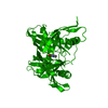
| ||||||||
| Unit cell |
|
-Components
| #1: Protein | Mass: 38201.625 Da / Num. of mol.: 1 Source method: isolated from a genetically manipulated source Source: (gene. exp.) Thermococcus litoralis (strain ATCC 51850 / DSM 5473 / JCM 8560 / NS-C) (archaea)Strain: ATCC 51850 / DSM 5473 / JCM 8560 / NS-C / Gene: OCC_00372 / Production host:  |
|---|---|
| #2: Chemical | ChemComp-PRO /   Type: L-peptide linking / Mass: 115.130 Da / Num. of mol.: 1 / Source method: obtained synthetically / Formula: C5H9NO2 / Feature type: SUBJECT OF INVESTIGATION Type: L-peptide linking / Mass: 115.130 Da / Num. of mol.: 1 / Source method: obtained synthetically / Formula: C5H9NO2 / Feature type: SUBJECT OF INVESTIGATION |
| #3: Water | ChemComp-HOH /  Mass: 18.015 Da / Num. of mol.: 29 / Source method: isolated from a natural source / Formula: H2O Mass: 18.015 Da / Num. of mol.: 29 / Source method: isolated from a natural source / Formula: H2O |
-Experimental details
-Experiment
| Experiment | Method: X-RAY DIFFRACTION / Number of used crystals: 1 |
|---|
- Sample preparation
| Crystal | Density Matthews: 4.35 Å3/Da / Density % sol: 71.72 % |
|---|---|
| Crystal grow | Temperature: 293 K / Method: vapor diffusion, sitting drop / pH: 7 Details: 0.1M HEPES pH 7.0, 30% (v/v) Jeffamine ED-2001 pH 7.0 |
-Data collection
| Diffraction | Mean temperature: 100 K / Serial crystal experiment: N |
|---|---|
| Diffraction source | Source: SYNCHROTRON / Site: SPring-8 / Beamline: BL38B1 / Wavelength: 1 Å |
| Detector | Type: MARMOSAIC 225 mm CCD / Detector: CCD / Date: Oct 8, 2017 |
| Radiation | Protocol: SINGLE WAVELENGTH / Monochromatic (M) / Laue (L): M / Scattering type: x-ray |
| Radiation wavelength | Wavelength: 1 Å / Relative weight: 1 |
| Reflection | Resolution: 2.7→50 Å / Num. obs: 18535 / % possible obs: 100 % / Redundancy: 20.1 % / Biso Wilson estimate: 58.24 Å2 / Rpim(I) all: 0.041 / Rrim(I) all: 0.185 / Rsym value: 0.18 / Net I/σ(I): 25.4 |
| Reflection shell | Resolution: 2.7→2.75 Å / Redundancy: 19.7 % / Mean I/σ(I) obs: 2 / Num. unique obs: 889 / CC1/2: 0.734 / Rpim(I) all: 0.446 / Rrim(I) all: 1.993 / Rsym value: 1.941 / % possible all: 100 |
- Processing
| Software |
| ||||||||||||||||||||||||||||||||||||||||||||||||||||||||||||||||||||||||||||||||||||
|---|---|---|---|---|---|---|---|---|---|---|---|---|---|---|---|---|---|---|---|---|---|---|---|---|---|---|---|---|---|---|---|---|---|---|---|---|---|---|---|---|---|---|---|---|---|---|---|---|---|---|---|---|---|---|---|---|---|---|---|---|---|---|---|---|---|---|---|---|---|---|---|---|---|---|---|---|---|---|---|---|---|---|---|---|---|
| Refinement | Method to determine structure: MOLECULAR REPLACEMENT Starting model: 1W61 Resolution: 2.7→46.793 Å / SU ML: 0.53 / Cross valid method: THROUGHOUT / σ(F): 1.34 / Phase error: 33.93
| ||||||||||||||||||||||||||||||||||||||||||||||||||||||||||||||||||||||||||||||||||||
| Solvent computation | Shrinkage radii: 0.9 Å / VDW probe radii: 1.11 Å | ||||||||||||||||||||||||||||||||||||||||||||||||||||||||||||||||||||||||||||||||||||
| Displacement parameters | Biso max: 98.65 Å2 / Biso mean: 58.1317 Å2 / Biso min: 30.76 Å2 | ||||||||||||||||||||||||||||||||||||||||||||||||||||||||||||||||||||||||||||||||||||
| Refinement step | Cycle: final / Resolution: 2.7→46.793 Å
| ||||||||||||||||||||||||||||||||||||||||||||||||||||||||||||||||||||||||||||||||||||
| Refine LS restraints |
| ||||||||||||||||||||||||||||||||||||||||||||||||||||||||||||||||||||||||||||||||||||
| LS refinement shell | Refine-ID: X-RAY DIFFRACTION / Rfactor Rfree error: 0 / Total num. of bins used: 13 / % reflection obs: 100 %
|
