 Movie
Movie Controller

[English] 日本語
 Yorodumi
- PDB-6hwt: Crystal structure of p38alpha in complex with a reduced photoswit... -
+ Open data

- Basic information
| Entry | Database: PDB / ID: 6hwt | |||||||||
|---|---|---|---|---|---|---|---|---|---|---|
| Title | Crystal structure of p38alpha in complex with a reduced photoswitchable 2-Azothiazol-based Inhibitor (compound 31) | |||||||||
 Components Components | Mitogen-activated protein kinase 14 MAPK14 MAPK14 | |||||||||
 Keywords Keywords | TRANSFERASE / p38alpha / MAPK14 / photoswitchable / Azothiazol | |||||||||
| Function / homology |  Function and homology information Function and homology informationpositive regulation of cyclase activity / Activation of PPARGC1A (PGC-1alpha) by phosphorylation / CD163 mediating an anti-inflammatory response / regulation of synaptic membrane adhesion / stress-induced premature senescence / cell surface receptor protein serine/threonine kinase signaling pathway / 3'-UTR-mediated mRNA stabilization / KSRP (KHSRP) binds and destabilizes mRNA / DSCAM interactions / cartilage condensation ...positive regulation of cyclase activity / Activation of PPARGC1A (PGC-1alpha) by phosphorylation / CD163 mediating an anti-inflammatory response / regulation of synaptic membrane adhesion / stress-induced premature senescence / cell surface receptor protein serine/threonine kinase signaling pathway / 3'-UTR-mediated mRNA stabilization / KSRP (KHSRP) binds and destabilizes mRNA / DSCAM interactions / cartilage condensation / cellular response to UV-B / Platelet sensitization by LDL / stress-activated protein kinase signaling cascade / mitogen-activated protein kinase p38 binding / positive regulation of myoblast fusion / positive regulation of muscle cell differentiation / negative regulation of hippo signaling / positive regulation of myotube differentiation / NFAT protein binding / Myogenesis / glucose import / response to dietary excess / Activation of the AP-1 family of transcription factors / ERK/MAPK targets / regulation of cytokine production involved in inflammatory response / p38MAPK cascade / fatty acid oxidation / cellular response to lipoteichoic acid / MAP kinase kinase activity / response to muramyl dipeptide / RHO GTPases Activate NADPH Oxidases / regulation of ossification / MAP kinase activity / mitogen-activated protein kinase / cellular response to vascular endothelial growth factor stimulus / positive regulation of myoblast differentiation / chondrocyte differentiation / vascular endothelial growth factor receptor signaling pathway / stress-activated MAPK cascade / skeletal muscle tissue development / signal transduction in response to DNA damage / lipopolysaccharide-mediated signaling pathway / negative regulation of inflammatory response to antigenic stimulus / positive regulation of cardiac muscle cell proliferation / p38MAPK events / striated muscle cell differentiation / response to muscle stretch / positive regulation of brown fat cell differentiation / positive regulation of interleukin-12 production / osteoclast differentiation / positive regulation of erythrocyte differentiation / placenta development / DNA damage checkpoint signaling / activated TAK1 mediates p38 MAPK activation / cellular response to ionizing radiation / stem cell differentiation / positive regulation of glucose import / negative regulation of canonical Wnt signaling pathway / response to insulin / NOD1/2 Signaling Pathway / bone development / cell morphogenesis / platelet activation / osteoblast differentiation / cellular response to virus / VEGFA-VEGFR2 Pathway / spindle pole / positive regulation of protein import into nucleus / ADP signalling through P2Y purinoceptor 1 / glucose metabolic process / positive regulation of reactive oxygen species metabolic process / chemotaxis / cellular senescence / cellular response to tumor necrosis factor / protein phosphatase binding / peptidyl-serine phosphorylation / angiogenesis / Oxidative Stress Induced Senescence / secretory granule lumen / cellular response to lipopolysaccharide / Regulation of TP53 Activity through Phosphorylation / ficolin-1-rich granule lumen / transcription by RNA polymerase II / cell surface receptor signaling pathway / intracellular signal transduction / nuclear speck / protein serine kinase activity / protein serine/threonine kinase activity / glutamatergic synapse / apoptotic process / Neutrophil degranulation / positive regulation of gene expression / regulation of transcription by RNA polymerase II / enzyme binding / signal transduction / positive regulation of transcription by RNA polymerase II / mitochondrion / extracellular region / nucleoplasm / ATP bindingSimilarity search - Function | |||||||||
| Biological species |  Homo sapiens (human) Homo sapiens (human) | |||||||||
| Method | X-RAY DIFFRACTION / SYNCHROTRON / MOLECULAR REPLACEMENT / Resolution: 1.7 Å | |||||||||
 Authors Authors | Mueller, M.P. / Rauh, D. | |||||||||
| Funding support |  Germany, 2items Germany, 2items
| |||||||||
 Citation Citation | Journal: Photochem. Photobiol. Sci. / Year: 2019 Title: 2-Azo-, 2-diazocine-thiazols and 2-azo-imidazoles as photoswitchable kinase inhibitors: limitations and pitfalls of the photoswitchable inhibitor approach. Authors: Schehr, M. / Ianes, C. / Weisner, J. / Heintze, L. / Muller, M.P. / Pichlo, C. / Charl, J. / Brunstein, E. / Ewert, J. / Lehr, M. / Baumann, U. / Rauh, D. / Knippschild, U. / Peifer, C. / Herges, R. | |||||||||
| History |
|
- Structure visualization
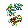
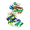
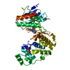
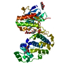
| Structure viewer | Molecule: MolmilJmol/JSmol |
|---|
- Downloads & links
-Download
| PDBx/mmCIF format | 6hwt.cif.gz | 159.4 KB | Display | PDBx/mmCIF format |
|---|---|---|---|---|
| PDB format | pdb6hwt.ent.gz | 123.1 KB | Display | PDB format |
| PDBx/mmJSON format | 6hwt.json.gz | Tree view | PDBx/mmJSON format | |
| Others |  Other downloads Other downloads |
-Validation report
| Arichive directory | https://data.pdbj.org/pub/pdb/validation_reports/hw/6hwtftp://data.pdbj.org/pub/pdb/validation_reports/hw/6hwt | HTTPS FTP |
|---|
-Related structure data
| Related structure data |  6hmpC  6hmrC  6hwuC  6hwvC 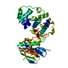 5n63S S: Starting model for refinement C: citing same article ( |
|---|---|
| Similar structure data |


-Links
 PDBj
PDBj


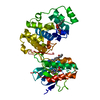
- Assembly
| Deposited unit | 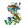
| ||||||||
|---|---|---|---|---|---|---|---|---|---|
| 1 |
| ||||||||
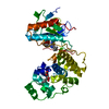
| Unit cell |
|
-Components
| #1: Protein | MAPK14 / MAPK 14 / Cytokine suppressive anti-inflammatory drug-binding protein / CSBP / MAP kinase MXI2 / ...MAPK 14 / Cytokine suppressive anti-inflammatory drug-binding protein / CSBP / MAP kinase MXI2 / MAX-interacting protein 2 / Mitogen-activated protein kinase p38 alpha / MAP kinase p38 alpha / Stress-activated protein kinase 2a / SAPK2a Mass: 41494.277 Da / Num. of mol.: 1 Source method: isolated from a genetically manipulated source Source: (gene. exp.) Homo sapiens (human) / Gene: MAPK14, CSBP, CSBP1, CSBP2, CSPB1, MXI2, SAPK2A / Production host:  Escherichia coli (E. coli) Escherichia coli (E. coli)References: UniProt: Q16539, mitogen-activated protein kinase | ||||
|---|---|---|---|---|---|
| #2: Sugar | Octyl glucoside  Type: D-saccharide / Mass: 292.369 Da / Num. of mol.: 3 / Source method: isolated from a natural source / Formula: C14H28O6 / Comment: detergent*YM Type: D-saccharide / Mass: 292.369 Da / Num. of mol.: 3 / Source method: isolated from a natural source / Formula: C14H28O6 / Comment: detergent*YM#3: Chemical | ChemComp-GXH / | 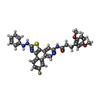  Mass: 569.649 Da / Num. of mol.: 1 / Source method: obtained synthetically / Formula: C31H28FN5O3S / Feature type: SUBJECT OF INVESTIGATION Mass: 569.649 Da / Num. of mol.: 1 / Source method: obtained synthetically / Formula: C31H28FN5O3S / Feature type: SUBJECT OF INVESTIGATION#4: Water | ChemComp-HOH / | Water Mass: 18.015 Da / Num. of mol.: 205 / Source method: isolated from a natural source / Formula: H2O Mass: 18.015 Da / Num. of mol.: 205 / Source method: isolated from a natural source / Formula: H2O |
-Experimental details
-Experiment
| Experiment | Method: X-RAY DIFFRACTION / Number of used crystals: 1 |
|---|
- Sample preparation
| Crystal | Density Matthews: 2.19 Å3/Da / Density % sol: 43.86 % |
|---|---|
| Crystal grow | Temperature: 293 K / Method: vapor diffusion, hanging drop / Details: 100 mM MES pH 5.6-6.2, 20-30 % PEG4000, 40mM BOG |
-Data collection
| Diffraction | Mean temperature: 100 K / Serial crystal experiment: N |
|---|---|
| Diffraction source | Source: SYNCHROTRON / Site: SLS  / Beamline: X10SA / Wavelength: 0.977928 Å / Beamline: X10SA / Wavelength: 0.977928 Å |
| Detector | Type: PSI PILATUS 6M / Detector: PIXEL / Date: Sep 8, 2017 |
| Radiation | Protocol: SINGLE WAVELENGTH / Monochromatic (M) / Laue (L): M / Scattering type: x-ray |
| Radiation wavelength | Wavelength: 0.977928 Å / Relative weight: 1 |
| Reflection | Resolution: 1.7→49.3 Å / Num. obs: 39982 / % possible obs: 98.5 % / Redundancy: 8.4 % / Rmerge(I) obs: 0.065 / Rrim(I) all: 0.069 / Net I/σ(I): 17 |
| Reflection shell | Resolution: 1.7→1.8 Å / Redundancy: 8.6 % / Rmerge(I) obs: 0.803 / Num. unique obs: 6261 / Rrim(I) all: 0.858 / % possible all: 100 |
- Processing
| Software |
| |||||||||||||||||||||||||||||||||||||||||||||||||||||||||||||||||||||||||||||||||||||||||||||||||||||||||
|---|---|---|---|---|---|---|---|---|---|---|---|---|---|---|---|---|---|---|---|---|---|---|---|---|---|---|---|---|---|---|---|---|---|---|---|---|---|---|---|---|---|---|---|---|---|---|---|---|---|---|---|---|---|---|---|---|---|---|---|---|---|---|---|---|---|---|---|---|---|---|---|---|---|---|---|---|---|---|---|---|---|---|---|---|---|---|---|---|---|---|---|---|---|---|---|---|---|---|---|---|---|---|---|---|---|---|
| Refinement | Method to determine structure: MOLECULAR REPLACEMENT Starting model: 5n63 Resolution: 1.7→49.257 Å / SU ML: 0.2 / Cross valid method: FREE R-VALUE / σ(F): 1.35 / Phase error: 28.93
| |||||||||||||||||||||||||||||||||||||||||||||||||||||||||||||||||||||||||||||||||||||||||||||||||||||||||
| Solvent computation | Shrinkage radii: 0.9 Å / VDW probe radii: 1.11 Å | |||||||||||||||||||||||||||||||||||||||||||||||||||||||||||||||||||||||||||||||||||||||||||||||||||||||||
| Refinement step | Cycle: LAST / Resolution: 1.7→49.257 Å
| |||||||||||||||||||||||||||||||||||||||||||||||||||||||||||||||||||||||||||||||||||||||||||||||||||||||||
| Refine LS restraints |
| |||||||||||||||||||||||||||||||||||||||||||||||||||||||||||||||||||||||||||||||||||||||||||||||||||||||||
| LS refinement shell |
| |||||||||||||||||||||||||||||||||||||||||||||||||||||||||||||||||||||||||||||||||||||||||||||||||||||||||
| Refinement TLS params. | S33: 0 Å ° / Method: refined / Refine-ID: X-RAY DIFFRACTION
| |||||||||||||||||||||||||||||||||||||||||||||||||||||||||||||||||||||||||||||||||||||||||||||||||||||||||
| Refinement TLS group |
|
