 ムービー
ムービー コントローラー


+ データを開く

- 基本情報
基本情報
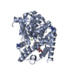
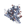
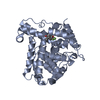
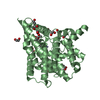
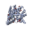
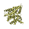
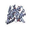
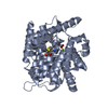
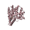
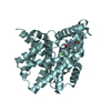
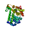
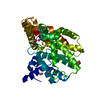
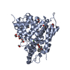
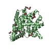
| 登録情報 | データベース: PDB / ID: 6fw3 | ||||||
|---|---|---|---|---|---|---|---|
| タイトル | Crystal structure of human phosphodiesterase 4D2 catalytic domain with inhibitor NPD-007 | ||||||
 要素 要素 | cAMP-specific 3',5'-cyclic phosphodiesterase 4D | ||||||
 キーワード キーワード | HYDROLASE / phosphodiesterase / cAMP hydrolysis / alternative splicing | ||||||
| 機能・相同性 |  機能・相同性情報 機能・相同性情報signaling receptor regulator activity / negative regulation of relaxation of cardiac muscle / negative regulation of heart contraction / 3',5'-cyclic-AMP phosphodiesterase / negative regulation of adenylate cyclase-activating G protein-coupled receptor signaling pathway / positive regulation of interleukin-5 production / regulation of cardiac muscle cell contraction / establishment of endothelial barrier / heterocyclic compound binding / beta-2 adrenergic receptor binding ...signaling receptor regulator activity / negative regulation of relaxation of cardiac muscle / negative regulation of heart contraction / 3',5'-cyclic-AMP phosphodiesterase / negative regulation of adenylate cyclase-activating G protein-coupled receptor signaling pathway / positive regulation of interleukin-5 production / regulation of cardiac muscle cell contraction / establishment of endothelial barrier / heterocyclic compound binding / beta-2 adrenergic receptor binding / regulation of calcium ion transmembrane transport via high voltage-gated calcium channel / voltage-gated calcium channel complex / adrenergic receptor signaling pathway / cAMP catabolic process / 3',5'-cyclic-nucleotide phosphodiesterase activity / regulation of cell communication by electrical coupling involved in cardiac conduction / 3',5'-cyclic-GMP phosphodiesterase activity / 3',5'-cyclic-AMP phosphodiesterase activity / DARPP-32 events / negative regulation of cAMP/PKA signal transduction / positive regulation of heart rate / cAMP binding / regulation of release of sequestered calcium ion into cytosol by sarcoplasmic reticulum / cellular response to epinephrine stimulus / calcium channel complex / positive regulation of interleukin-2 production / regulation of heart rate / cellular response to cAMP / Turbulent (oscillatory, disturbed) flow shear stress activates signaling by PIEZO1 and integrins in endothelial cells / calcium channel regulator activity / positive regulation of type II interferon production / T cell receptor signaling pathway / ATPase binding / scaffold protein binding / nuclear membrane / G alpha (s) signalling events / transmembrane transporter binding / apical plasma membrane / cilium / centrosome / enzyme binding / nucleoplasm / metal ion binding / membrane / plasma membrane / cytosol 類似検索 - 分子機能 | ||||||
| 生物種 |  Homo sapiens (ヒト) Homo sapiens (ヒト) | ||||||
| 手法 |  X線回折 / シンクロトロン / 分子置換 / 解像度: 1.78 Å X線回折 / シンクロトロン / 分子置換 / 解像度: 1.78 Å | ||||||
 データ登録者 データ登録者 | Singh, A.K. / Brown, D.G. | ||||||
| 資金援助 | 1件
| ||||||
 引用 引用 | ジャーナル: To be published タイトル: hPDE4D2 structure with inhibitor NPD-007 著者: Salado, I.G. / Moreno, C. / Sakaine, G. / Singh, A.K. / Blaazer, A.R. / Siderius, M. / Matheeussen, A. / Gul, S. / Maes, L. / Leurs, R. / Brown, D.G. / Augustyns, K. | ||||||
| 履歴 |
|
- 構造の表示
構造の表示
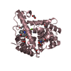
| 構造ビューア | 分子: MolmilJmol/JSmol |
|---|
- ダウンロードとリンク
-ダウンロード
| PDBx/mmCIF形式 | 6fw3.cif.gz | 312.3 KB | 表示 | PDBx/mmCIF形式 |
|---|---|---|---|---|
| PDB形式 | pdb6fw3.ent.gz | 250.2 KB | 表示 | PDB形式 |
| PDBx/mmJSON形式 | 6fw3.json.gz | ツリー表示 | PDBx/mmJSON形式 | |
| その他 |  その他のダウンロード その他のダウンロード |
-検証レポート
| アーカイブディレクトリ | https://data.pdbj.org/pub/pdb/validation_reports/fw/6fw3ftp://data.pdbj.org/pub/pdb/validation_reports/fw/6fw3 | HTTPS FTP |
|---|
-関連構造データ
| 関連構造データ | 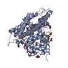 3sl3S S: 精密化の開始モデル |
|---|---|
| 類似構造データ |


-リンク
 PDBj
PDBj


- 集合体
集合体
| 登録構造単位 | 
| ||||||||
|---|---|---|---|---|---|---|---|---|---|
| 1 | 
| ||||||||
| 2 | 
| ||||||||
| 3 | 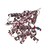
| ||||||||
| 4 | 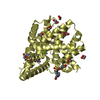
| ||||||||
| 単位格子 |
|
-要素
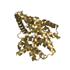
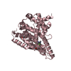
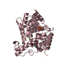
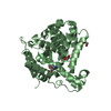
-タンパク質 , 1種, 4分子 ABCD
| #1: タンパク質 | 分子量: 41808.242 Da / 分子数: 4 / 由来タイプ: 組換発現 / 詳細: residues 381-740 / 由来: (組換発現) Homo sapiens (ヒト) / 遺伝子: PDE4D, DPDE3 / プラスミド: pET15b / 発現宿主:  |
|---|
-非ポリマー , 8種, 1020分子 


| #2: 化合物 | ChemComp-ZN /  分子量: 65.409 Da / 分子数: 4 / 由来タイプ: 合成 / 式: Zn 分子量: 65.409 Da / 分子数: 4 / 由来タイプ: 合成 / 式: Zn#3: 化合物 | ChemComp-MG /  分子量: 24.305 Da / 分子数: 4 / 由来タイプ: 合成 / 式: Mg 分子量: 24.305 Da / 分子数: 4 / 由来タイプ: 合成 / 式: Mg#4: 化合物 | ChemComp-EDO /  分子量: 62.068 Da / 分子数: 59 / 由来タイプ: 合成 / 式: C2H6O2 分子量: 62.068 Da / 分子数: 59 / 由来タイプ: 合成 / 式: C2H6O2#5: 化合物 |  分子量: 106.120 Da / 分子数: 2 / 由来タイプ: 合成 / 式: C4H10O3 分子量: 106.120 Da / 分子数: 2 / 由来タイプ: 合成 / 式: C4H10O3#6: 化合物 | ChemComp-EPE /  分子量: 238.305 Da / 分子数: 5 / 由来タイプ: 合成 / 式: C8H18N2O4S / コメント: pH緩衝剤*YM 分子量: 238.305 Da / 分子数: 5 / 由来タイプ: 合成 / 式: C8H18N2O4S / コメント: pH緩衝剤*YM#7: 化合物 | ChemComp-E8H /  分子量: 508.566 Da / 分子数: 4 / 由来タイプ: 合成 / 式: C27H32N4O6 / タイプ: SUBJECT OF INVESTIGATION 分子量: 508.566 Da / 分子数: 4 / 由来タイプ: 合成 / 式: C27H32N4O6 / タイプ: SUBJECT OF INVESTIGATION#8: 化合物 | ChemComp-PG4 / |  分子量: 194.226 Da / 分子数: 1 / 由来タイプ: 合成 / 式: C8H18O5 / コメント: 沈殿剤*YM 分子量: 194.226 Da / 分子数: 1 / 由来タイプ: 合成 / 式: C8H18O5 / コメント: 沈殿剤*YM#9: 水 | ChemComp-HOH / | 分子量: 18.015 Da / 分子数: 941 / 由来タイプ: 天然 / 式: H2O |
|---|
-実験情報
-実験
| 実験 | 手法: X線回折 / 使用した結晶の数: 1 |
|---|
- 試料調製
試料調製
| 結晶 | マシュー密度: 2.65 Å3/Da / 溶媒含有率: 53.65 % |
|---|---|
| 結晶化 | 温度: 293 K / 手法: 蒸気拡散法, ハンギングドロップ法 / pH: 7.5 / 詳細: 24% PEG 3350, 30% Ethylene Glycol, 0.1 M HEPES |
-データ収集
| 回折 | 平均測定温度: 100 K |
|---|---|
| 放射光源 | 由来: シンクロトロン / サイト: Diamond  / ビームライン: I04-1 / 波長: 0.91587 Å / ビームライン: I04-1 / 波長: 0.91587 Å |
| 検出器 | タイプ: DECTRIS PILATUS 6M-F / 検出器: PIXEL / 日付: 2018年2月16日 |
| 放射 | プロトコル: SINGLE WAVELENGTH / 単色(M)・ラウエ(L): M / 散乱光タイプ: x-ray |
| 放射波長 | 波長: 0.91587 Å / 相対比: 1 |
| 反射 | 解像度: 1.78→65.23 Å / Num. obs: 170039 / % possible obs: 100 % / 冗長度: 6.4 % / CC1/2: 0.999 / Rmerge(I) obs: 0.061 / Rpim(I) all: 0.026 / Rrim(I) all: 0.067 / Net I/σ(I): 15 |
| 反射 シェル | 解像度: 1.78→1.83 Å / 冗長度: 5.5 % / Rmerge(I) obs: 0.684 / Mean I/σ(I) obs: 2 / Num. unique obs: 12471 / CC1/2: 0.725 / Rpim(I) all: 0.323 / Rrim(I) all: 0.759 / % possible all: 100 |
- 解析
解析
| ソフトウェア |
| ||||||||||||||||||||||||||||||||||||||||||||||||||||||||||||||||||||||||||||||||||||||||||||||||||||||||||||||||||||||||||||||||||||||||||||||||||||||||||||||||||||||||||||||||||||||
|---|---|---|---|---|---|---|---|---|---|---|---|---|---|---|---|---|---|---|---|---|---|---|---|---|---|---|---|---|---|---|---|---|---|---|---|---|---|---|---|---|---|---|---|---|---|---|---|---|---|---|---|---|---|---|---|---|---|---|---|---|---|---|---|---|---|---|---|---|---|---|---|---|---|---|---|---|---|---|---|---|---|---|---|---|---|---|---|---|---|---|---|---|---|---|---|---|---|---|---|---|---|---|---|---|---|---|---|---|---|---|---|---|---|---|---|---|---|---|---|---|---|---|---|---|---|---|---|---|---|---|---|---|---|---|---|---|---|---|---|---|---|---|---|---|---|---|---|---|---|---|---|---|---|---|---|---|---|---|---|---|---|---|---|---|---|---|---|---|---|---|---|---|---|---|---|---|---|---|---|---|---|---|---|
| 精密化 | 構造決定の手法: 分子置換 開始モデル: 3SL3 解像度: 1.78→65.23 Å / Cor.coef. Fo:Fc: 0.967 / Cor.coef. Fo:Fc free: 0.95 / SU B: 2.704 / SU ML: 0.08 / 交差検証法: THROUGHOUT / ESU R: 0.105 / ESU R Free: 0.106 / 詳細: HYDROGENS HAVE BEEN ADDED IN THE RIDING POSITIONS
| ||||||||||||||||||||||||||||||||||||||||||||||||||||||||||||||||||||||||||||||||||||||||||||||||||||||||||||||||||||||||||||||||||||||||||||||||||||||||||||||||||||||||||||||||||||||
| 溶媒の処理 | イオンプローブ半径: 0.8 Å / 減衰半径: 0.8 Å / VDWプローブ半径: 1.2 Å | ||||||||||||||||||||||||||||||||||||||||||||||||||||||||||||||||||||||||||||||||||||||||||||||||||||||||||||||||||||||||||||||||||||||||||||||||||||||||||||||||||||||||||||||||||||||
| 原子変位パラメータ | Biso mean: 31.367 Å2
| ||||||||||||||||||||||||||||||||||||||||||||||||||||||||||||||||||||||||||||||||||||||||||||||||||||||||||||||||||||||||||||||||||||||||||||||||||||||||||||||||||||||||||||||||||||||
| 精密化ステップ | サイクル: 1 / 解像度: 1.78→65.23 Å
| ||||||||||||||||||||||||||||||||||||||||||||||||||||||||||||||||||||||||||||||||||||||||||||||||||||||||||||||||||||||||||||||||||||||||||||||||||||||||||||||||||||||||||||||||||||||
| 拘束条件 |
|