 Movie
Movie Controller

[English] 日本語
 Yorodumi
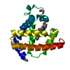
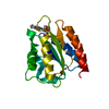
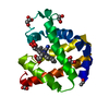
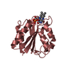
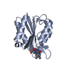
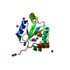
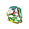
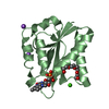
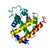
- PDB-6fsg: Crystal structure of oxidised Flavodoxin 1 from Bacillus cereus (... -
+ Open data

- Basic information
| Entry | Database: PDB / ID: 6fsg | ||||||
|---|---|---|---|---|---|---|---|
| Title | Crystal structure of oxidised Flavodoxin 1 from Bacillus cereus (1.27 A resolution) | ||||||
 Components Components | Flavodoxin | ||||||
 Keywords Keywords | ELECTRON TRANSPORT / flavodoxin / electron transfer / FMN | ||||||
| Function / homology |  Function and homology information Function and homology informationoxidoreductase activity, acting on NAD(P)H / FMN binding / electron transfer activity Similarity search - Function | ||||||
| Biological species |  | ||||||
| Method |  X-RAY DIFFRACTION / SYNCHROTRON / MOLECULAR REPLACEMENT / molecular replacement / Resolution: 1.27 Å X-RAY DIFFRACTION / SYNCHROTRON / MOLECULAR REPLACEMENT / molecular replacement / Resolution: 1.27 Å | ||||||
 Authors Authors | Gudim, I. / Lofstad, M. / Hersleth, H.-P. | ||||||
| Funding support |  Norway, 1items Norway, 1items
| ||||||
 Citation Citation | Journal: Protein Sci. / Year: 2018 Title: High-resolution crystal structures reveal a mixture of conformers of the Gly61-Asp62 peptide bond in an oxidized flavodoxin from Bacillus cereus. Authors: Gudim, I. / Lofstad, M. / van Beek, W. / Hersleth, H.P. | ||||||
| History |
|
- Structure visualization
| Structure viewer | Molecule: MolmilJmol/JSmol |
|---|
- Downloads & links
-Download
| PDBx/mmCIF format | 6fsg.cif.gz | 112 KB | Display | PDBx/mmCIF format |
|---|---|---|---|---|
| PDB format | pdb6fsg.ent.gz | 87 KB | Display | PDB format |
| PDBx/mmJSON format | 6fsg.json.gz | Tree view | PDBx/mmJSON format | |
| Others |  Other downloads Other downloads |
-Validation report
| Summary document | 6fsg_validation.pdf.gz | 788.8 KB | Display | wwPDB validaton report |
|---|---|---|---|---|
| Full document | 6fsg_full_validation.pdf.gz | 791.8 KB | Display | |
| Data in XML | 6fsg_validation.xml.gz | 10.9 KB | Display | |
| Data in CIF | 6fsg_validation.cif.gz | 16.2 KB | Display | |
| Arichive directory | https://data.pdbj.org/pub/pdb/validation_reports/fs/6fsgftp://data.pdbj.org/pub/pdb/validation_reports/fs/6fsg | HTTPS FTP |
-Related structure data
| Related structure data | 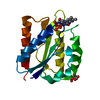 6fsiC 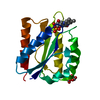 6ft1C 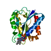 1wsbS S: Starting model for refinement C: citing same article ( |
|---|---|
| Similar structure data |
-Links
 PDBj
PDBj


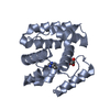
- Assembly
| Deposited unit | 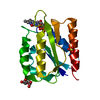
| ||||||||
|---|---|---|---|---|---|---|---|---|---|
| 1 |
| ||||||||
| Unit cell |
| ||||||||
| Components on special symmetry positions |
|
-Components
| #1: Protein | Mass: 16115.039 Da / Num. of mol.: 1 Source method: isolated from a genetically manipulated source Source: (gene. exp.) Gene: BC_1376 / Plasmid: pET22b(+) / Production host: |
|---|---|
| #2: Chemical | ChemComp-FMN /   Mass: 456.344 Da / Num. of mol.: 1 / Source method: obtained synthetically / Formula: C17H21N4O9P Mass: 456.344 Da / Num. of mol.: 1 / Source method: obtained synthetically / Formula: C17H21N4O9P |
| #3: Chemical | ChemComp-SO4 /   Mass: 96.063 Da / Num. of mol.: 1 / Source method: obtained synthetically / Formula: SO4 Mass: 96.063 Da / Num. of mol.: 1 / Source method: obtained synthetically / Formula: SO4 |
| #4: Sugar | ChemComp-GLC /   Type: D-saccharide, alpha linking / Mass: 180.156 Da / Num. of mol.: 1 Type: D-saccharide, alpha linking / Mass: 180.156 Da / Num. of mol.: 1Source method: isolated from a genetically manipulated source Formula: C6H12O6 |
| #5: Water | ChemComp-HOH /  Mass: 18.015 Da / Num. of mol.: 217 / Source method: isolated from a natural source / Formula: H2O Mass: 18.015 Da / Num. of mol.: 217 / Source method: isolated from a natural source / Formula: H2O |
-Experimental details
-Experiment
| Experiment | Method: X-RAY DIFFRACTION / Number of used crystals: 1 |
|---|
- Sample preparation
| Crystal | Density Matthews: 2.25 Å3/Da / Density % sol: 45.26 % |
|---|---|
| Crystal grow | Temperature: 298 K / Method: vapor diffusion, sitting drop / pH: 5 Details: 3.6 M ammonium sulfate and 0.1 M citric acid pH 4 (final pH 5) |
-Data collection
| Diffraction | Mean temperature: 100 K |
|---|---|
| Diffraction source | Source: SYNCHROTRON / Site: ESRF  / Beamline: BM1A / Wavelength: 0.6983 Å / Beamline: BM1A / Wavelength: 0.6983 Å |
| Detector | Type: MAR scanner 345 mm plate / Detector: IMAGE PLATE / Date: Jun 3, 2011 |
| Radiation | Protocol: SINGLE WAVELENGTH / Monochromatic (M) / Laue (L): M / Scattering type: x-ray |
| Radiation wavelength | Wavelength: 0.6983 Å / Relative weight: 1 |
| Reflection | Resolution: 1.27→35.2 Å / Num. obs: 38867 / % possible obs: 99.3 % / Redundancy: 5.2 % / Biso Wilson estimate: 6.54 Å2 / CC1/2: 0.998 / Rmerge(I) obs: 0.05 / Rpim(I) all: 0.023 / Rrim(I) all: 0.055 / Net I/σ(I): 19.7 |
| Reflection shell | Resolution: 1.27→1.29 Å / Redundancy: 3.6 % / Rmerge(I) obs: 0.073 / Mean I/σ(I) obs: 9.8 / Num. unique obs: 1722 / CC1/2: 0.992 / Rpim(I) all: 0.042 / Rrim(I) all: 0.085 / % possible all: 90.8 |
-Phasing
| Phasing | Method: molecular replacement |
|---|
- Processing
| Software |
| |||||||||||||||||||||||||||||||||||||||||||||||||||||||||||||||||||||||||||||||||||||||||||||||||||||||||
|---|---|---|---|---|---|---|---|---|---|---|---|---|---|---|---|---|---|---|---|---|---|---|---|---|---|---|---|---|---|---|---|---|---|---|---|---|---|---|---|---|---|---|---|---|---|---|---|---|---|---|---|---|---|---|---|---|---|---|---|---|---|---|---|---|---|---|---|---|---|---|---|---|---|---|---|---|---|---|---|---|---|---|---|---|---|---|---|---|---|---|---|---|---|---|---|---|---|---|---|---|---|---|---|---|---|---|
| Refinement | Method to determine structure: MOLECULAR REPLACEMENT Starting model: 1WSB Resolution: 1.27→35.195 Å / SU ML: 0.08 / Cross valid method: THROUGHOUT / σ(F): 1.34 / Phase error: 15.63
| |||||||||||||||||||||||||||||||||||||||||||||||||||||||||||||||||||||||||||||||||||||||||||||||||||||||||
| Solvent computation | Shrinkage radii: 0.9 Å / VDW probe radii: 1.11 Å | |||||||||||||||||||||||||||||||||||||||||||||||||||||||||||||||||||||||||||||||||||||||||||||||||||||||||
| Displacement parameters | Biso max: 55.76 Å2 / Biso mean: 9.6792 Å2 / Biso min: 2.64 Å2 | |||||||||||||||||||||||||||||||||||||||||||||||||||||||||||||||||||||||||||||||||||||||||||||||||||||||||
| Refinement step | Cycle: final / Resolution: 1.27→35.195 Å
| |||||||||||||||||||||||||||||||||||||||||||||||||||||||||||||||||||||||||||||||||||||||||||||||||||||||||
| LS refinement shell | Refine-ID: X-RAY DIFFRACTION / Rfactor Rfree error: 0 / Total num. of bins used: 14
|
