 Movie
Movie Controller

[English] 日本語
 Yorodumi
- PDB-6exy: Neutron crystal structure of perdeuterated galectin-3C in complex... -
+ Open data

- Basic information
| Entry | Database: PDB / ID: 6exy | ||||||
|---|---|---|---|---|---|---|---|
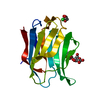
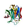
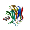
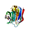
| Title | Neutron crystal structure of perdeuterated galectin-3C in complex with glycerol | ||||||
 Components Components | Galectin-3 | ||||||
 Keywords Keywords | SUGAR BINDING PROTEIN / galectin / hydrogen bonding / molecular recognition / perdeuteration | ||||||
| Function / homology |  Function and homology information Function and homology informationnegative regulation of protein tyrosine phosphatase activity / negative regulation of immunological synapse formation / RUNX2 regulates genes involved in differentiation of myeloid cells / negative regulation of T cell activation via T cell receptor contact with antigen bound to MHC molecule on antigen presenting cell / regulation of T cell apoptotic process / mononuclear cell migration / IgE binding / positive regulation of mononuclear cell migration / negative regulation of endocytosis / eosinophil chemotaxis ...negative regulation of protein tyrosine phosphatase activity / negative regulation of immunological synapse formation / RUNX2 regulates genes involved in differentiation of myeloid cells / negative regulation of T cell activation via T cell receptor contact with antigen bound to MHC molecule on antigen presenting cell / regulation of T cell apoptotic process / mononuclear cell migration / IgE binding / positive regulation of mononuclear cell migration / negative regulation of endocytosis / eosinophil chemotaxis / regulation of extrinsic apoptotic signaling pathway via death domain receptors / RUNX1 regulates transcription of genes involved in differentiation of myeloid cells / protein phosphatase inhibitor activity / negative regulation of T cell receptor signaling pathway / positive chemotaxis / macrophage chemotaxis / regulation of T cell proliferation / positive regulation of calcium ion import / chemoattractant activity / monocyte chemotaxis / Advanced glycosylation endproduct receptor signaling / ficolin-1-rich granule membrane / immunological synapse / laminin binding / epithelial cell differentiation / molecular condensate scaffold activity / neutrophil chemotaxis / RNA splicing / secretory granule membrane / positive regulation of protein-containing complex assembly / negative regulation of extrinsic apoptotic signaling pathway / positive regulation of protein localization to plasma membrane / spliceosomal complex / mRNA processing / carbohydrate binding / collagen-containing extracellular matrix / protein phosphatase binding / mitochondrial inner membrane / innate immune response / Neutrophil degranulation / cell surface / extracellular space / RNA binding / extracellular exosome / extracellular region / nucleoplasm / membrane / nucleus / plasma membrane / cytosol / cytoplasmSimilarity search - Function | ||||||
| Biological species |  Homo sapiens (human) Homo sapiens (human) | ||||||
| Method | X-RAY DIFFRACTION / NEUTRON DIFFRACTION / SYNCHROTRON / NUCLEAR REACTOR / FOURIER SYNTHESIS / Resolution: 1.1 Å | ||||||
 Authors Authors | Manzoni, F. / Schrader, T.E. / Ostermann, A. / Oksanen, E. / Logan, D.T. | ||||||
| Funding support |  Sweden, 1items Sweden, 1items
| ||||||
 Citation Citation | Journal: J. Med. Chem. / Year: 2018 Title: Elucidation of Hydrogen Bonding Patterns in Ligand-Free, Lactose- and Glycerol-Bound Galectin-3C by Neutron Crystallography to Guide Drug Design. Authors: Manzoni, F. / Wallerstein, J. / Schrader, T.E. / Ostermann, A. / Coates, L. / Akke, M. / Blakeley, M.P. / Oksanen, E. / Logan, D.T. #1: Journal: Acta Crystallogr D Struct Biol / Year: 2016 Title: Perdeuteration, crystallization, data collection and comparison of five neutron diffraction data sets of complexes of human galectin-3C. Authors: Manzoni, F. / Saraboji, K. / Sprenger, J. / Kumar, R. / Noresson, A.L. / Nilsson, U.J. / Leffler, H. / Fisher, S.Z. / Schrader, T.E. / Ostermann, A. / Coates, L. / Blakeley, M.P. / Oksanen, E. / Logan, D.T. | ||||||
| History |
|
- Structure visualization
| Structure viewer | Molecule: MolmilJmol/JSmol |
|---|
- Downloads & links
-Download
| PDBx/mmCIF format | 6exy.cif.gz | 165.2 KB | Display | PDBx/mmCIF format |
|---|---|---|---|---|
| PDB format | pdb6exy.ent.gz | 116.9 KB | Display | PDB format |
| PDBx/mmJSON format | 6exy.json.gz | Tree view | PDBx/mmJSON format | |
| Others |  Other downloads Other downloads |
-Validation report
| Arichive directory | https://data.pdbj.org/pub/pdb/validation_reports/ex/6exyftp://data.pdbj.org/pub/pdb/validation_reports/ex/6exy | HTTPS FTP |
|---|
-Related structure data
| Related structure data |  6eymC  6f2qC  3zsjS S: Starting model for refinement C: citing same article ( |
|---|---|
| Similar structure data |


-Links
 PDBj
PDBj


- Assembly
| Deposited unit | 
| ||||||||||
|---|---|---|---|---|---|---|---|---|---|---|---|
| 1 |
| ||||||||||
| Unit cell |
|
-Components
| #1: Protein | / Gal-3 / 35 kDa lectin / Carbohydrate-binding protein 35 / CBP 35 / Galactose-specific lectin 3 / ...Gal-3 / 35 kDa lectin / Carbohydrate-binding protein 35 / CBP 35 / Galactose-specific lectin 3 / Galactoside-binding protein / GALBP / IgE-binding protein / L-31 / Laminin-binding protein / Lectin L-29 / Mac-2 antigen Mass: 15832.244 Da / Num. of mol.: 1 Source method: isolated from a genetically manipulated source Source: (gene. exp.) Homo sapiens (human) / Gene: LGALS3, MAC2 / Production host:  Escherichia coli (E. coli) / References: UniProt: P17931 Escherichia coli (E. coli) / References: UniProt: P17931 |
|---|---|
| #2: Chemical | ChemComp-GOL / Glycerol  Mass: 92.094 Da / Num. of mol.: 1 / Source method: obtained synthetically / Formula: C3H8O3 Mass: 92.094 Da / Num. of mol.: 1 / Source method: obtained synthetically / Formula: C3H8O3 |
| #3: Water | ChemComp-HOH / Water Mass: 18.015 Da / Num. of mol.: 101 / Source method: isolated from a natural source / Formula: H2O Mass: 18.015 Da / Num. of mol.: 101 / Source method: isolated from a natural source / Formula: H2O |
-Experimental details
-Experiment
| Experiment |
|
|---|
- Sample preparation
| Crystal | Density Matthews: 2.19 Å3/Da / Density % sol: 44 % / Description: Volume approximately 1.0 mm3 |
|---|---|
| Crystal grow | Temperature: 293 K / Method: vapor diffusion, sitting drop / pH: 7.5 Details: 12-15% PEG 4000 OR PEG 3000, 0.1M MGCL2, 0.015M BETA MERCAPTOETHANOL, 0.1M TRIS-DCL, PD 7.9, 0.4M NaSCN. All dissolved in D2O. Crystal grown in a 15 + 15 microlitre sitting drop that was ...Details: 12-15% PEG 4000 OR PEG 3000, 0.1M MGCL2, 0.015M BETA MERCAPTOETHANOL, 0.1M TRIS-DCL, PD 7.9, 0.4M NaSCN. All dissolved in D2O. Crystal grown in a 15 + 15 microlitre sitting drop that was first equilibrated for 1 week. A small crystal grown at 20-28% PEG was introduced. The drop was fed with fresh protein by adding 3-4 micro litres of protein with 10 mM lactose every 3-4 days for 3 months. Then the lactose was exchanged for glycerol by dialysis for at least one month against 10% glycerol (1.37 M), 24% PEG 4000 in the same buffer. For details see Manzoni et al. (2016). |
-Data collection
| Diffraction |
| |||||||||||||||||||||||||||
|---|---|---|---|---|---|---|---|---|---|---|---|---|---|---|---|---|---|---|---|---|---|---|---|---|---|---|---|---|
| Diffraction source |
| |||||||||||||||||||||||||||
| Detector |
| |||||||||||||||||||||||||||
| Radiation |
| |||||||||||||||||||||||||||
| Radiation wavelength |
| |||||||||||||||||||||||||||
| Reflection | Biso Wilson estimate: 11.45 Å2 / Entry-ID: 6EXY
| |||||||||||||||||||||||||||
| Reflection shell |
|
- Processing
| Software |
| |||||||||||||||||||||||||||||||||||||||||||||||||||||||||||||||||||||||||||||||||||||||||||||||||||||||||||||||||||||||||||||||||||||||||||||||||||
|---|---|---|---|---|---|---|---|---|---|---|---|---|---|---|---|---|---|---|---|---|---|---|---|---|---|---|---|---|---|---|---|---|---|---|---|---|---|---|---|---|---|---|---|---|---|---|---|---|---|---|---|---|---|---|---|---|---|---|---|---|---|---|---|---|---|---|---|---|---|---|---|---|---|---|---|---|---|---|---|---|---|---|---|---|---|---|---|---|---|---|---|---|---|---|---|---|---|---|---|---|---|---|---|---|---|---|---|---|---|---|---|---|---|---|---|---|---|---|---|---|---|---|---|---|---|---|---|---|---|---|---|---|---|---|---|---|---|---|---|---|---|---|---|---|---|---|---|---|
| Refinement | SU ML: 0.0845 / Cross valid method: FREE R-VALUE / Method to determine structure
| |||||||||||||||||||||||||||||||||||||||||||||||||||||||||||||||||||||||||||||||||||||||||||||||||||||||||||||||||||||||||||||||||||||||||||||||||||
| Refinement step | Cycle: LAST / Resolution: 1.1→31.93 Å
| |||||||||||||||||||||||||||||||||||||||||||||||||||||||||||||||||||||||||||||||||||||||||||||||||||||||||||||||||||||||||||||||||||||||||||||||||||
| Refine LS restraints |
| |||||||||||||||||||||||||||||||||||||||||||||||||||||||||||||||||||||||||||||||||||||||||||||||||||||||||||||||||||||||||||||||||||||||||||||||||||
| LS refinement shell |
|
