Protocol: SINGLE WAVELENGTH / Monochromatic (M) / Laue (L): M / Scattering type: x-ray
Radiation wavelength
Wavelength: 1 Å / Relative weight: 1
Reflection
Resolution: 1.75→50 Å / Num. obs: 160030 / % possible obs: 99.3 % / Redundancy: 3.7 % / Rmerge(I) obs: 0.055 / Net I/σ(I): 24.7
Reflection shell
Resolution: 1.75→1.81 Å / Redundancy: 3.1 % / Rmerge(I) obs: 0.247 / Mean I/σ(I) obs: 4.9 / % possible all: 94.4
-
Processing
Software
Name
Version
Classification
REFMAC
5.8.0107
refinement
HKL-2000
datareduction
HKL-2000
datascaling
PHENIX
phasing
Refinement
Method to determine structure: SAD / Resolution: 1.75→28.3 Å / Cor.coef. Fo:Fc: 0.95 / Cor.coef. Fo:Fc free: 0.94 / Cross valid method: THROUGHOUT / ESU R: 0.107 / ESU R Free: 0.096 / Details: HYDROGENS HAVE BEEN ADDED IN THE RIDING POSITIONS
Rfactor
Num. reflection
% reflection
Selection details
Rfree
0.19184
7983
5 %
RANDOM
Rwork
0.17658
-
-
-
obs
0.17734
151942
99.29 %
-
Solvent computation
Ion probe radii: 0.8 Å / Shrinkage radii: 0.8 Å / VDW probe radii: 1.2 Å
 Movie
Movie Controller

 Yorodumi
 Open data
 Basic information
 Components
 Keywords
 Function and homology information
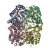
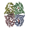
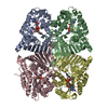
 Paracoccus laeviglucosivorans Nakamura 2015 (bacteria)
 X-RAY DIFFRACTION /
  Authors
 Japan, 1items
  Citation
 Structure visualization
 Downloads & links
 Other downloads


 PDBj
PDBj Assembly

 Mass: 59.044 Da / Num. of mol.: 4 / Source method: obtained synthetically / Formula: C2H3O2
 Mass: 18.015 Da / Num. of mol.: 852 / Source method: isolated from a natural source / Formula: H2O
 Sample preparation
 Processing