 Movie
Movie Controller

[English] 日本語
 Yorodumi
- PDB-5qh4: PanDDA analysis group deposition of models with modelled events (... -
+ Open data

- Basic information
| Entry | Database: PDB / ID: 5qh4 | ||||||
|---|---|---|---|---|---|---|---|
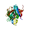
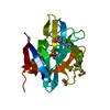
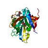
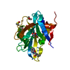
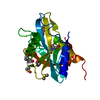
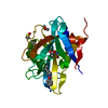
| Title | PanDDA analysis group deposition of models with modelled events (e.g. bound ligands) -- Crystal Structure of NUDT7 in complex with NUOOA000220a | ||||||
 Components Components | Peroxisomal coenzyme A diphosphatase NUDT7 | ||||||
 Keywords Keywords | HYDROLASE / PanDDA / SGC - Diamond I04-1 fragment screening / NUDIX domain / XChemExplorer | ||||||
| Function / homology |  Function and homology information Function and homology informationbutyryl-CoA catabolic process / propionyl-CoA metabolic process / propionyl-CoA catabolic process / coenzyme A diphosphatase / Peroxisomal lipid metabolism / malonyl-CoA catabolic process / coenzyme A diphosphatase activity / medium-chain fatty-acyl-CoA catabolic process / coenzyme A catabolic process / acetyl-CoA catabolic process ...butyryl-CoA catabolic process / propionyl-CoA metabolic process / propionyl-CoA catabolic process / coenzyme A diphosphatase / Peroxisomal lipid metabolism / malonyl-CoA catabolic process / coenzyme A diphosphatase activity / medium-chain fatty-acyl-CoA catabolic process / coenzyme A catabolic process / acetyl-CoA catabolic process / succinyl-CoA catabolic process / nucleoside diphosphate metabolic process / snoRNA binding / peroxisomal matrix / Hydrolases; Acting on acid anhydrides; In phosphorus-containing anhydrides / brown fat cell differentiation / Peroxisomal protein import / peroxisome / manganese ion binding / magnesium ion binding / cytosol Similarity search - Function | ||||||
| Biological species |  Homo sapiens (human) Homo sapiens (human) | ||||||
| Method |  X-RAY DIFFRACTION / SYNCHROTRON / FOURIER SYNTHESIS / molecular replacement / Resolution: 1.67 Å X-RAY DIFFRACTION / SYNCHROTRON / FOURIER SYNTHESIS / molecular replacement / Resolution: 1.67 Å | ||||||
 Authors Authors | Krojer, T. / Talon, R. / Fairhead, M. / Diaz Saez, L. / Bradley, A.R. / Aimon, A. / Collins, P. / Brandao-Neto, J. / Douangamath, A. / Ruda, G.F. ...Krojer, T. / Talon, R. / Fairhead, M. / Diaz Saez, L. / Bradley, A.R. / Aimon, A. / Collins, P. / Brandao-Neto, J. / Douangamath, A. / Ruda, G.F. / Szommer, T. / Srikannathasan, V. / Elkins, J. / Spencer, J. / London, N. / Nelson, A. / Brennan, P.E. / Huber, K. / Bountra, C. / Arrowsmith, C.H. / Edwards, A. / von Delft, F. | ||||||
 Citation Citation | Journal: To Be Published Title: PanDDA analysis group deposition of models with modelled events (e.g. bound ligands) Authors: Krojer, T. / Talon, R. / Fairhead, M. / Diaz Saez, L. / Bradley, A.R. / Aimon, A. / Collins, P. / Brandao-Neto, J. / Douangamath, A. / Ruda, G.F. / Szommer, T. / Srikannathasan, V. / Elkins, ...Authors: Krojer, T. / Talon, R. / Fairhead, M. / Diaz Saez, L. / Bradley, A.R. / Aimon, A. / Collins, P. / Brandao-Neto, J. / Douangamath, A. / Ruda, G.F. / Szommer, T. / Srikannathasan, V. / Elkins, J. / Spencer, J. / London, N. / Nelson, A. / Brennan, P.E. / Huber, K. / Bountra, C. / Arrowsmith, C.H. / Edwards, A. / von Delft, F. | ||||||
| History |
|
- Structure visualization
| Structure viewer | Molecule: MolmilJmol/JSmol |
|---|
- Downloads & links
-Download
| PDBx/mmCIF format | 5qh4.cif.gz | 57.6 KB | Display | PDBx/mmCIF format |
|---|---|---|---|---|
| PDB format | pdb5qh4.ent.gz | 40.1 KB | Display | PDB format |
| PDBx/mmJSON format | 5qh4.json.gz | Tree view | PDBx/mmJSON format | |
| Others |  Other downloads Other downloads |
-Validation report
| Arichive directory | https://data.pdbj.org/pub/pdb/validation_reports/qh/5qh4ftp://data.pdbj.org/pub/pdb/validation_reports/qh/5qh4 | HTTPS FTP |
|---|
-Group deposition
| ID | G_1002045 (37 entries) |
|---|---|
| Title | PanDDA analysis group deposition of models with modelled events (e.g. bound ligands) |
| Type | changed state |
| Description | human NUDT7 screened against the 3D-Fragment Consortium Library by X-ray Crystallography at the XChem facility of Diamond Light Source beamline I04-1 |


-Related structure data
| Related structure data |  5t3pS S: Starting model for refinement |
|---|---|
| Similar structure data | |
| Experimental dataset #1 | Data reference: 10.5281/zenodo.1244111 / Data set type: other data / Details: Complete PanDDA analysis / Metadata reference: 10.5281/zenodo.1244111 |


-Links
 PDBj
PDBj

- Assembly
| Deposited unit | 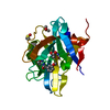
| ||||||||
|---|---|---|---|---|---|---|---|---|---|
| 1 |
| ||||||||
| Unit cell |
|
-Components
| #1: Protein | Mass: 22197.600 Da / Num. of mol.: 1 Source method: isolated from a genetically manipulated source Source: (gene. exp.) Homo sapiens (human) / Gene: NUDT7 / Production host:  References: UniProt: P0C024, Hydrolases; Acting on acid anhydrides; In phosphorus-containing anhydrides | ||||||
|---|---|---|---|---|---|---|---|
| #2: Chemical |   Mass: 59.044 Da / Num. of mol.: 2 / Source method: obtained synthetically / Formula: C2H3O2 Mass: 59.044 Da / Num. of mol.: 2 / Source method: obtained synthetically / Formula: C2H3O2#3: Chemical |   Mass: 78.133 Da / Num. of mol.: 2 / Source method: obtained synthetically / Formula: C2H6OS / Comment: DMSO, precipitant*YM Mass: 78.133 Da / Num. of mol.: 2 / Source method: obtained synthetically / Formula: C2H6OS / Comment: DMSO, precipitant*YM#4: Chemical | ChemComp-H47 / |   Mass: 232.235 Da / Num. of mol.: 1 / Source method: obtained synthetically / Formula: C12H12N2O3 Mass: 232.235 Da / Num. of mol.: 1 / Source method: obtained synthetically / Formula: C12H12N2O3#5: Water | ChemComp-HOH / |  Mass: 18.015 Da / Num. of mol.: 159 / Source method: isolated from a natural source / Formula: H2O Mass: 18.015 Da / Num. of mol.: 159 / Source method: isolated from a natural source / Formula: H2O |
-Experimental details
-Experiment
| Experiment | Method: X-RAY DIFFRACTION / Number of used crystals: 1 |
|---|
- Sample preparation
| Crystal | Density Matthews: 4.16 Å3/Da / Density % sol: 70.41 % / Mosaicity: 0.07 ° |
|---|---|
| Crystal grow | Temperature: 293 K / Method: vapor diffusion, sitting drop / pH: 5.5 Details: 0.1M bis-tris pH 5.5 -- 0.1M ammonium acetate -- 5%(w/v) PEG10K |
-Data collection
| Diffraction | Mean temperature: 100 K | ||||||||||||||||||||||||||||||
|---|---|---|---|---|---|---|---|---|---|---|---|---|---|---|---|---|---|---|---|---|---|---|---|---|---|---|---|---|---|---|---|
| Diffraction source | Source: SYNCHROTRON / Site: Diamond  / Beamline: I04-1 / Wavelength: 0.91587 Å / Beamline: I04-1 / Wavelength: 0.91587 Å | ||||||||||||||||||||||||||||||
| Detector | Type: DECTRIS PILATUS 6M / Detector: PIXEL / Date: Sep 7, 2017 | ||||||||||||||||||||||||||||||
| Radiation | Protocol: SINGLE WAVELENGTH / Scattering type: x-ray | ||||||||||||||||||||||||||||||
| Radiation wavelength | Wavelength: 0.91587 Å / Relative weight: 1 | ||||||||||||||||||||||||||||||
| Reflection | Resolution: 1.67→28.98 Å / Num. obs: 42885 / % possible obs: 99.8 % / Redundancy: 10.1 % / CC1/2: 1 / Rmerge(I) obs: 0.043 / Rpim(I) all: 0.014 / Rrim(I) all: 0.046 / Net I/σ(I): 29.3 / Num. measured all: 431624 / Scaling rejects: 0 | ||||||||||||||||||||||||||||||
| Reflection shell | Diffraction-ID: 1
|
-Phasing
| Phasing | Method: molecular replacement |
|---|
- Processing
| Software |
| |||||||||||||||||||||||||||||||||||||||||||||||||||||||||||||||||||||||||||
|---|---|---|---|---|---|---|---|---|---|---|---|---|---|---|---|---|---|---|---|---|---|---|---|---|---|---|---|---|---|---|---|---|---|---|---|---|---|---|---|---|---|---|---|---|---|---|---|---|---|---|---|---|---|---|---|---|---|---|---|---|---|---|---|---|---|---|---|---|---|---|---|---|---|---|---|---|
| Refinement | Method to determine structure: FOURIER SYNTHESIS Starting model: 5T3P Resolution: 1.67→107.93 Å / Cor.coef. Fo:Fc: 0.957 / Cor.coef. Fo:Fc free: 0.948 / SU B: 1.59 / SU ML: 0.053 / Cross valid method: THROUGHOUT / σ(F): 0 / ESU R: 0.076 / ESU R Free: 0.075 / Stereochemistry target values: MAXIMUM LIKELIHOOD Details: HYDROGENS HAVE BEEN ADDED IN THE RIDING POSITIONS U VALUES : REFINED INDIVIDUALLY
| |||||||||||||||||||||||||||||||||||||||||||||||||||||||||||||||||||||||||||
| Solvent computation | Ion probe radii: 0.8 Å / Shrinkage radii: 0.8 Å / VDW probe radii: 1.2 Å / Solvent model: MASK | |||||||||||||||||||||||||||||||||||||||||||||||||||||||||||||||||||||||||||
| Displacement parameters | Biso max: 92.45 Å2 / Biso mean: 29.51 Å2 / Biso min: 15.57 Å2
| |||||||||||||||||||||||||||||||||||||||||||||||||||||||||||||||||||||||||||
| Refinement step | Cycle: final / Resolution: 1.67→107.93 Å
| |||||||||||||||||||||||||||||||||||||||||||||||||||||||||||||||||||||||||||
| Refine LS restraints |
| |||||||||||||||||||||||||||||||||||||||||||||||||||||||||||||||||||||||||||
| LS refinement shell | Resolution: 1.668→1.711 Å / Total num. of bins used: 20
|
