 Movie
Movie Controller

[English] 日本語
 Yorodumi
- PDB-5pb8: PanDDA analysis group deposition -- Crystal Structure of BAZ2B in... -
+ Open data

- Basic information
| Entry | Database: PDB / ID: 5pb8 | ||||||
|---|---|---|---|---|---|---|---|
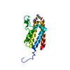
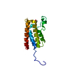
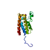
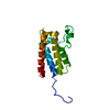
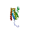
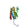
| Title | PanDDA analysis group deposition -- Crystal Structure of BAZ2B in complex with N09522a | ||||||
 Components Components | Bromodomain adjacent to zinc finger domain protein 2B | ||||||
 Keywords Keywords |  DNA BINDING PROTEIN / PanDDA / SGC - Diamond I04-1 fragment screening / bromodomain / epigenetics DNA BINDING PROTEIN / PanDDA / SGC - Diamond I04-1 fragment screening / bromodomain / epigenetics | ||||||
| Function / homology |  Function and homology informationchromatin remodeling / regulation of transcription by RNA polymerase II / DNA binding / metal ion binding / nucleus Function and homology informationchromatin remodeling / regulation of transcription by RNA polymerase II / DNA binding / metal ion binding / nucleusSimilarity search - Function | ||||||
| Biological species |  Homo sapiens (human) Homo sapiens (human) | ||||||
| Method | X-RAY DIFFRACTION / SYNCHROTRON / FOURIER SYNTHESIS / molecular replacement / Resolution: 1.649 Å | ||||||
 Authors Authors | Pearce, N.M. / Krojer, T. / Talon, R. / Bradley, A.R. / Fairhead, M. / Sethi, R. / Wright, N. / MacLean, E. / Collins, P. / Brandao-Neto, J. ...Pearce, N.M. / Krojer, T. / Talon, R. / Bradley, A.R. / Fairhead, M. / Sethi, R. / Wright, N. / MacLean, E. / Collins, P. / Brandao-Neto, J. / Douangamath, A. / Renjie, Z. / Dias, A. / Vollmar, M. / Ng, J. / Brennan, P.E. / Cox, O. / Bountra, C. / Arrowsmith, C.H. / Edwards, A. / von Delft, F. | ||||||
 Citation Citation | Journal: Nat Commun / Year: 2017 Title: A multi-crystal method for extracting obscured crystallographic states from conventionally uninterpretable electron density. Authors: Pearce, N.M. / Krojer, T. / Bradley, A.R. / Collins, P. / Nowak, R.P. / Talon, R. / Marsden, B.D. / Kelm, S. / Shi, J. / Deane, C.M. / von Delft, F. | ||||||
| History |
|
- Structure visualization
| Structure viewer | Molecule: MolmilJmol/JSmol |
|---|
- Downloads & links
-Download
| PDBx/mmCIF format | 5pb8.cif.gz | 44.7 KB | Display | PDBx/mmCIF format |
|---|---|---|---|---|
| PDB format | pdb5pb8.ent.gz | 30.5 KB | Display | PDB format |
| PDBx/mmJSON format | 5pb8.json.gz | Tree view | PDBx/mmJSON format | |
| Others |  Other downloads Other downloads |
-Validation report
| Arichive directory | https://data.pdbj.org/pub/pdb/validation_reports/pb/5pb8ftp://data.pdbj.org/pub/pdb/validation_reports/pb/5pb8 | HTTPS FTP |
|---|
-Group deposition
| ID | G_1002018 (9 entries) |
|---|---|
| Title | PanDDA analysis group deposition of models with modelled events (e.g. bound ligands) |
| Type | changed state |
| Description | bromodomain of human BAZ2B screened against the ZENOBIA Fragment Library by X-ray Crystallography at the XChem facility of Diamond Light Source beamline I04-1. Check out the PanDDA event maps at https://zenodo.org/record/290199/files/0_index.html |


-Related structure data
| Related structure data |  3g0lS S: Starting model for refinement |
|---|---|
| Similar structure data |


-Links
 PDBj
PDBj
- Assembly
| Deposited unit | 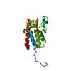
| ||||||||
|---|---|---|---|---|---|---|---|---|---|
| 1 |
| ||||||||
| Unit cell |
| ||||||||
| Components on special symmetry positions |
|
-Components
| #1: Protein | Mass: 16090.326 Da / Num. of mol.: 1 Source method: isolated from a genetically manipulated source Source: (gene. exp.) Homo sapiens (human) / Gene: BAZ2B, KIAA1476 / Production host:  escherichia coli (E. coli) / References: UniProt: Q9UIF8 escherichia coli (E. coli) / References: UniProt: Q9UIF8 | ||||||
|---|---|---|---|---|---|---|---|
| #2: Chemical | ChemComp-EDO / Ethylene glycol  Mass: 62.068 Da / Num. of mol.: 4 / Source method: obtained synthetically / Formula: C2H6O2 Mass: 62.068 Da / Num. of mol.: 4 / Source method: obtained synthetically / Formula: C2H6O2#3: Chemical | ChemComp-AC6 / | Piceol  Mass: 136.148 Da / Num. of mol.: 1 / Source method: obtained synthetically / Formula: C8H8O2 Mass: 136.148 Da / Num. of mol.: 1 / Source method: obtained synthetically / Formula: C8H8O2#4: Water | ChemComp-HOH / | Water Mass: 18.015 Da / Num. of mol.: 201 / Source method: isolated from a natural source / Formula: H2O Mass: 18.015 Da / Num. of mol.: 201 / Source method: isolated from a natural source / Formula: H2ONonpolymer details | yes CC(c1ccc(cc1)O)=O None 12.98 39.25 39.25 CC(c1ccc(cc1)O)=O 4 - High Confidence None 0.77 34. ... | |
-Experimental details
-Experiment
| Experiment | Method: X-RAY DIFFRACTION / Number of used crystals: 1 |
|---|
- Sample preparation
| Crystal | Density Matthews: 3.59 Å3/Da / Density % sol: 65.74 % / Mosaicity: 0.28 ° |
|---|---|
| Crystal grow | Temperature: 277 K / Method: vapor diffusion, sitting drop / pH: 7 / Details: 30% PEG600 -- 0.1M MES pH 6.0 |
-Data collection
| Diffraction | Mean temperature: 100 K | ||||||||||||||||||||||||||||||
|---|---|---|---|---|---|---|---|---|---|---|---|---|---|---|---|---|---|---|---|---|---|---|---|---|---|---|---|---|---|---|---|
| Diffraction source | Source: SYNCHROTRON / Site: Diamond  / Beamline: I04-1 / Wavelength: 0.92 Å / Beamline: I04-1 / Wavelength: 0.92 Å | ||||||||||||||||||||||||||||||
| Detector | Type: DECTRIS PILATUS 2M / Detector: PIXEL / Date: Mar 10, 2013 | ||||||||||||||||||||||||||||||
| Radiation | Protocol: SINGLE WAVELENGTH / Scattering type: x-ray | ||||||||||||||||||||||||||||||
| Radiation wavelength | Wavelength: 0.92 Å / Relative weight: 1 | ||||||||||||||||||||||||||||||
| Reflection | Resolution: 1.65→29.07 Å / Num. obs: 27756 / % possible obs: 98.2 % / Redundancy: 6.4 % / Biso Wilson estimate: 25.24 Å2 / CC1/2: 0.999 / Rmerge(I) obs: 0.066 / Rpim(I) all: 0.028 / Rrim(I) all: 0.072 / Net I/σ(I): 15.1 / Num. measured all: 178006 / Scaling rejects: 0 | ||||||||||||||||||||||||||||||
| Reflection shell | Diffraction-ID: 1
|
-Phasing
| Phasing | Method: molecular replacement |
|---|
- Processing
| Software |
| |||||||||||||||||||||||||||||||||||||||||||||||||||||||||||||||||||||||||||||
|---|---|---|---|---|---|---|---|---|---|---|---|---|---|---|---|---|---|---|---|---|---|---|---|---|---|---|---|---|---|---|---|---|---|---|---|---|---|---|---|---|---|---|---|---|---|---|---|---|---|---|---|---|---|---|---|---|---|---|---|---|---|---|---|---|---|---|---|---|---|---|---|---|---|---|---|---|---|---|
| Refinement | Method to determine structure: FOURIER SYNTHESIS Starting model: 3G0L Resolution: 1.649→29.07 Å / SU ML: 0.22 / Cross valid method: THROUGHOUT / σ(F): 1.34 / Phase error: 22.89 / Stereochemistry target values: ML
| |||||||||||||||||||||||||||||||||||||||||||||||||||||||||||||||||||||||||||||
| Solvent computation | Shrinkage radii: 0.9 Å / VDW probe radii: 1.11 Å / Solvent model: FLAT BULK SOLVENT MODEL | |||||||||||||||||||||||||||||||||||||||||||||||||||||||||||||||||||||||||||||
| Displacement parameters | Biso max: 84.38 Å2 / Biso mean: 31.2733 Å2 / Biso min: 15.51 Å2 | |||||||||||||||||||||||||||||||||||||||||||||||||||||||||||||||||||||||||||||
| Refinement step | Cycle: final / Resolution: 1.649→29.07 Å
| |||||||||||||||||||||||||||||||||||||||||||||||||||||||||||||||||||||||||||||
| Refine LS restraints |
| |||||||||||||||||||||||||||||||||||||||||||||||||||||||||||||||||||||||||||||
| LS refinement shell | Refine-ID: X-RAY DIFFRACTION / Total num. of bins used: 10
|
