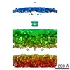
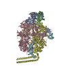
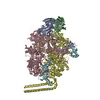
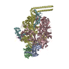
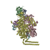
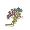
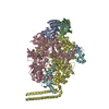
Journal: Elife / Year: 2017 Title: Structure and organisation of the archaellum machinery. Authors: Bertram Daum / Janet Vonck / Annett Bellack / Paushali Chaudhury / Robert Reichelt / Sonja-Verena Albers / Reinhard Rachel / Werner Kühlbrandt / Abstract: The archaellum is the macromolecular machinery that Archaea use for propulsion or surface adhesion, enabling them to proliferate and invade new territories. The molecular composition of the ...The archaellum is the macromolecular machinery that Archaea use for propulsion or surface adhesion, enabling them to proliferate and invade new territories. The molecular composition of the archaellum and of the motor that drives it appears to be entirely distinct from that of the functionally equivalent bacterial flagellum and flagellar motor. Yet, the structure of the archaellum machinery is scarcely known. Using combined modes of electron cryo-microscopy (cryoEM), we have solved the structure of the archaellum filament at 4.2 Å resolution and visualise the architecture and organisation of its motor complex . This allows us to build a structural model combining the archaellum and its motor complex, paving the way to a molecular understanding of archaeal swimming motion.
Average exposure time: 8 sec. / Electron dose: 60 e/Å2 / Detector mode: COUNTING / Film or detector model: GATAN K2 SUMMIT (4k x 4k) / Num. of real images: 297
In the structure databanks used in Yorodumi, some data are registered as the other names, "COVID-19 virus" and "2019-nCoV". Here are the details of the virus and the list of structure data.
Jan 31, 2019. EMDB accession codes are about to change! (news from PDBe EMDB page)
EMDB accession codes are about to change! (news from PDBe EMDB page)
The allocation of 4 digits for EMDB accession codes will soon come to an end. Whilst these codes will remain in use, new EMDB accession codes will include an additional digit and will expand incrementally as the available range of codes is exhausted. The current 4-digit format prefixed with “EMD-” (i.e. EMD-XXXX) will advance to a 5-digit format (i.e. EMD-XXXXX), and so on. It is currently estimated that the 4-digit codes will be depleted around Spring 2019, at which point the 5-digit format will come into force.
The EM Navigator/Yorodumi systems omit the EMD- prefix.
Related info.:Q: What is EMD? / ID/Accession-code notation in Yorodumi/EM Navigator
Yorodumi is a browser for structure data from EMDB, PDB, SASBDB, etc.
This page is also the successor to EM Navigator detail page, and also detail information page/front-end page for Omokage search.
The word "yorodu" (or yorozu) is an old Japanese word meaning "ten thousand". "mi" (miru) is to see.
Related info.:EMDB / PDB / SASBDB / Comparison of 3 databanks / Yorodumi Search / Aug 31, 2016. New EM Navigator & Yorodumi / Yorodumi Papers / Jmol/JSmol / Function and homology information / Changes in new EM Navigator and Yorodumi
 Movie
Movie Controller


 Open data
 Basic information
 Components
 Keywords
 Function and homology information
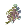
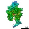
 Pyrococcus furiosus DSM 3638 (archaea)
 Authors
 Citation

 Structure visualization
 Downloads & links
 Other downloads
 PDBj
PDBj Assembly
 Sample preparation
 Electron microscopy imaging
 FIELD EMISSION GUN / Accelerating voltage: 300 kV / Illumination mode: FLOOD BEAM
 Processing
