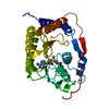
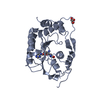
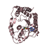
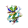
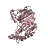
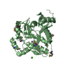
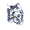
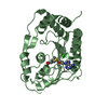
[English] 日本語
Yorodumi
- PDB-5njv: Flavivirus NS5 domain -

+
Open data


ID or keywords:

Loading...

-
Basic information

Entry
Database: PDB / ID: 5njv
TitleFlavivirus NS5 domain
ComponentsNS5
KeywordsVIRAL PROTEIN / Zika / flavivirus / NS5 / SAH
Function / homology
Function and homology information


symbiont-mediated suppression of host JAK-STAT cascade via inhibition of host TYK2 activity / flavivirin / symbiont-mediated suppression of host JAK-STAT cascade via inhibition of STAT2 activity / symbiont-mediated suppression of host JAK-STAT cascade via inhibition of STAT1 activity / negative regulation of innate immune response / ribonucleoside triphosphate phosphatase activity / viral capsid / nucleoside-triphosphate phosphatase / double-stranded RNA binding / 4 iron, 4 sulfur cluster binding ...symbiont-mediated suppression of host JAK-STAT cascade via inhibition of host TYK2 activity / flavivirin / symbiont-mediated suppression of host JAK-STAT cascade via inhibition of STAT2 activity / symbiont-mediated suppression of host JAK-STAT cascade via inhibition of STAT1 activity / negative regulation of innate immune response / ribonucleoside triphosphate phosphatase activity / viral capsid / nucleoside-triphosphate phosphatase / double-stranded RNA binding / 4 iron, 4 sulfur cluster binding / mRNA (guanine-N7)-methyltransferase / methyltransferase cap1 / clathrin-dependent endocytosis of virus by host cell / mRNA (nucleoside-2'-O-)-methyltransferase activity / mRNA 5'-cap (guanine-N7-)-methyltransferase activity / RNA helicase activity / host cell endoplasmic reticulum membrane / host cell perinuclear region of cytoplasm / protein dimerization activity / molecular adaptor activity / RNA helicase / induction by virus of host autophagy / symbiont entry into host cell / RNA-directed RNA polymerase / viral RNA genome replication / RNA-dependent RNA polymerase activity / serine-type endopeptidase activity / fusion of virus membrane with host endosome membrane / centrosome / viral envelope / lipid binding / symbiont-mediated suppression of host type I interferon-mediated signaling pathway / host cell nucleus / structural molecule activity / virion attachment to host cell / GTP binding / virion membrane / ATP hydrolysis activity / proteolysis / extracellular region / ATP binding / membrane / metal ion binding
Similarity search - Function
: / : / Flavivirus envelope glycoprotein E, stem/anchor domain / RNA-directed RNA polymerase, thumb domain, Flavivirus / Flavivirus RNA-directed RNA polymerase, thumb domain / Flavivirus capsid protein C superfamily / Flavivirus non-structural protein NS2B / Flavivirus NS3 helicase, C-terminal helical domain / Genome polyprotein, Flavivirus / Flavivirus non-structural protein NS4A ...: / : / Flavivirus envelope glycoprotein E, stem/anchor domain / RNA-directed RNA polymerase, thumb domain, Flavivirus / Flavivirus RNA-directed RNA polymerase, thumb domain / Flavivirus capsid protein C superfamily / Flavivirus non-structural protein NS2B / Flavivirus NS3 helicase, C-terminal helical domain / Genome polyprotein, Flavivirus / Flavivirus non-structural protein NS4A / Flavivirus non-structural protein NS2B / Flavivirus capsid protein C / Flavivirus non-structural protein NS4B / mRNA cap 0/1 methyltransferase / Flavivirus capsid protein C / Flavivirus non-structural protein NS4B / Flavivirus non-structural protein NS4A / Flavivirus NS2B domain profile. / mRNA cap 0 and cap 1 methyltransferase (EC 2.1.1.56 and EC 2.1.1.57) domain profile. / Flavivirus non-structural protein NS2A / Flavivirus non-structural protein NS2A / Flavivirus NS3, petidase S7 / Peptidase S7, Flavivirus NS3 serine protease / Flavivirus NS3 protease (NS3pro) domain profile. / Envelope glycoprotein M, flavivirus / Flavivirus envelope glycoprotein M / RNA-directed RNA polymerase, flavivirus / Flavivirus RNA-directed RNA polymerase, fingers and palm domains / Flavivirus non-structural Protein NS1 / Flavivirus non-structural protein NS1 / Envelope glycoprotein M superfamily, flavivirus / Flavivirus polyprotein propeptide / Flavivirus polyprotein propeptide superfamily / Flavivirus polyprotein propeptide / Flaviviral glycoprotein E, central domain, subdomain 1 / Flaviviral glycoprotein E, central domain, subdomain 2 / Flavivirus envelope glycoprotein E, Stem/Anchor domain / Flavivirus glycoprotein E, immunoglobulin-like domain / Flavivirus envelope glycoprotein E, Stem/Anchor domain superfamily / Flavivirus glycoprotein, immunoglobulin-like domain / Flavivirus glycoprotein central and dimerisation domain / Flavivirus glycoprotein, central and dimerisation domains / Ribosomal RNA methyltransferase, FtsJ domain / FtsJ-like methyltransferase / Flavivirus/Alphavirus glycoprotein, immunoglobulin-like domain superfamily / Flavivirus glycoprotein, central and dimerisation domain superfamily / Flaviviral glycoprotein E, dimerisation domain / DEAD box, Flavivirus / Flavivirus DEAD domain / Vaccinia Virus protein VP39 / helicase superfamily c-terminal domain / Immunoglobulin E-set / Superfamilies 1 and 2 helicase C-terminal domain profile. / Superfamilies 1 and 2 helicase ATP-binding type-1 domain profile. / DEAD-like helicases superfamily / Helicase, C-terminal / Helicase superfamily 1/2, ATP-binding domain / RNA-directed RNA polymerase, catalytic domain / RdRp of positive ssRNA viruses catalytic domain profile. / S-adenosyl-L-methionine-dependent methyltransferase superfamily / Peptidase S1, PA clan / DNA/RNA polymerase superfamily / P-loop containing nucleoside triphosphate hydrolase / Rossmann fold / 3-Layer(aba) Sandwich / Alpha Beta
Similarity search - Domain/homology
S-ADENOSYLMETHIONINE / Genome polyprotein / Genome polyprotein
Similarity search - Component
Biological speciesZika virus
MethodX-RAY DIFFRACTION / SYNCHROTRON / MOLECULAR REPLACEMENT / Resolution: 2 Å
AuthorsTalapatra, S.K. / Chatrin, C. / Kozielski, F.
CitationJournal: Oncotarget / Year: 2018
Title: The structure of the binary methyltransferase-SAH complex from Zika virus reveals a novel conformation for the mechanism of mRNA capping.
Authors: Chatrin, C. / Talapatra, S.K. / Canard, B. / Kozielski, F.
History
DepositionMar 29, 2017Deposition site: PDBE / Processing site: PDBE
Revision 1.0Jan 24, 2018Provider: repository / Type: Initial release
Revision 1.1Apr 18, 2018Group: Data collection / Database references / Category: citation / Item: _citation.pdbx_database_id_PubMed / _citation.title
Revision 1.2Jan 17, 2024Group: Data collection / Database references ...Data collection / Database references / Derived calculations / Refinement description
Category: chem_comp_atom / chem_comp_bond ...chem_comp_atom / chem_comp_bond / database_2 / pdbx_initial_refinement_model / struct_conn
Item: _database_2.pdbx_DOI / _database_2.pdbx_database_accession ..._database_2.pdbx_DOI / _database_2.pdbx_database_accession / _struct_conn.pdbx_dist_value / _struct_conn.ptnr1_auth_asym_id / _struct_conn.ptnr1_label_asym_id / _struct_conn.ptnr2_auth_asym_id / _struct_conn.ptnr2_label_asym_id / _struct_conn.ptnr2_symmetry

-
Structure visualization

Structure viewerMolecule:
MolmilJmol/JSmol

Downloads & links

-
Assembly

Deposited unit
A: NS5
B: NS5
C: NS5
D: NS5
hetero molecules


Theoretical massNumber of molelcules
Total (without water)118,04912
Polymers116,3134
Non-polymers1,7368
Water10,791599
1
A: NS5
hetero molecules


Theoretical massNumber of molelcules
Total (without water)29,5484
Polymers29,0781
Non-polymers4693
Water181
TypeNameSymmetry operationNumber
identity operation1_555x,y,z1
MethodPISA
2
B: NS5
hetero molecules


Theoretical massNumber of molelcules
Total (without water)29,5484
Polymers29,0781
Non-polymers4693
Water181
TypeNameSymmetry operationNumber
identity operation1_555x,y,z1
MethodPISA
3
C: NS5
hetero molecules


Theoretical massNumber of molelcules
Total (without water)29,4772
Polymers29,0781
Non-polymers3981
Water181
TypeNameSymmetry operationNumber
identity operation1_555x,y,z1
MethodPISA
4
D: NS5
hetero molecules


Theoretical massNumber of molelcules
Total (without water)29,4772
Polymers29,0781
Non-polymers3981
Water181
TypeNameSymmetry operationNumber
identity operation1_555x,y,z1
MethodPISA
Unit cell
Length a, b, c (Å)125.920, 125.920, 125.854
Angle α, β, γ (deg.)90.00, 90.00, 120.00
Int Tables number169
Space group name H-MP61

-
Components

#1: Protein
NS5


Mass: 29078.240 Da / Num. of mol.: 4 / Fragment: UNP residues 2524-2785
Source method: isolated from a genetically manipulated source
Source: (gene. exp.) Zika virus (strain Mr 766) / Production host: Escherichia coli (E. coli) / References: UniProt: A0A146CJG7, UniProt: A0A024B7W1*PLUS
#2: Chemical
ChemComp-SAM / S-ADENOSYLMETHIONINE / S-Adenosyl methionine


Mass: 398.437 Da / Num. of mol.: 4 / Source method: obtained synthetically / Formula: C15H22N6O5S
#3: Chemical
ChemComp-CL / CHLORIDE ION / Chloride


Mass: 35.453 Da / Num. of mol.: 4 / Source method: obtained synthetically / Formula: Cl
#4: Water ChemComp-HOH / water / Water


Mass: 18.015 Da / Num. of mol.: 599 / Source method: isolated from a natural source / Formula: H2O

-
Experimental details

-
Experiment

ExperimentMethod: X-RAY DIFFRACTION / Number of used crystals: 1

-
Sample preparation

CrystalDensity Matthews: 2.4 Å3/Da / Density % sol: 48.79 %
Crystal growTemperature: 291 K / Method: vapor diffusion, hanging drop / pH: 7 / Details: 21% PEG 400 0.1 M Tris, pH 7.0

-
Data collection

DiffractionMean temperature: 77 K
Diffraction sourceSource: SYNCHROTRON / Site: Diamond / Beamline: I04-1 / Wavelength: 0.92 Å
DetectorType: DECTRIS PILATUS 6M-F / Detector: PIXEL / Date: Feb 4, 2017
RadiationProtocol: SINGLE WAVELENGTH / Monochromatic (M) / Laue (L): M / Scattering type: x-ray
Radiation wavelengthWavelength: 0.92 Å / Relative weight: 1
ReflectionResolution: 2→54.53 Å / Num. obs: 76301 / % possible obs: 100 % / Redundancy: 7.9 % / Rmerge(I) obs: 0.082 / Net I/σ(I): 15.8
Reflection shellResolution: 2→2.07 Å / Redundancy: 7.6 % / Rmerge(I) obs: 0.63 / Mean I/σ(I) obs: 2.4 / % possible all: 100

-
Processing

Software
NameVersionClassification
PHENIX(1.10.1_2155: ???)refinement
iMOSFLMdata reduction
SCALAdata scaling
PHASERphasing
RefinementMethod to determine structure: MOLECULAR REPLACEMENT
Starting model: 5KQR
Resolution: 2→54.53 Å / SU ML: 0.31 / Cross valid method: FREE R-VALUE / σ(F): 1.37 / Phase error: 29.01
RfactorNum. reflection% reflection
Rfree0.258 3741 4.91 %
Rwork0.201 --
obs0.204 76215 100 %
Solvent computationShrinkage radii: 0.9 Å / VDW probe radii: 1.11 Å
Refinement stepCycle: LAST / Resolution: 2→54.53 Å
ProteinNucleic acidLigandSolventTotal
Num. atoms7899 0 112 599 8610
Refine LS restraints
Refine-IDTypeDev idealNumber
X-RAY DIFFRACTIONf_bond_d0.0128242
X-RAY DIFFRACTIONf_angle_d1.19711163
X-RAY DIFFRACTIONf_dihedral_angle_d27.084900
X-RAY DIFFRACTIONf_chiral_restr0.0641217
X-RAY DIFFRACTIONf_plane_restr0.0071417
LS refinement shell
Resolution (Å)Rfactor RfreeNum. reflection RfreeRfactor RworkNum. reflection RworkRefine-ID% reflection obs (%)
2.0001-2.02540.38321180.31882682X-RAY DIFFRACTION100
2.0254-2.05210.35781390.30062631X-RAY DIFFRACTION100
2.0521-2.08020.33121520.27332686X-RAY DIFFRACTION100
2.0802-2.10990.32651690.2672631X-RAY DIFFRACTION100
2.1099-2.14140.34621500.25292710X-RAY DIFFRACTION100
2.1414-2.17480.33151270.25872647X-RAY DIFFRACTION100
2.1748-2.21050.32351490.24682695X-RAY DIFFRACTION100
2.2105-2.24860.29471330.24362695X-RAY DIFFRACTION100
2.2486-2.28950.30681280.23772706X-RAY DIFFRACTION100
2.2895-2.33360.29191260.22512646X-RAY DIFFRACTION100
2.3336-2.38120.30271340.21562690X-RAY DIFFRACTION100
2.3812-2.4330.27831160.21882678X-RAY DIFFRACTION100
2.433-2.48960.32431390.21442698X-RAY DIFFRACTION100
2.4896-2.55180.32321420.2262654X-RAY DIFFRACTION100
2.5518-2.62080.27361210.22522741X-RAY DIFFRACTION100
2.6208-2.69790.32381250.21832685X-RAY DIFFRACTION100
2.6979-2.7850.24581520.20042683X-RAY DIFFRACTION100
2.785-2.88460.27271410.1942663X-RAY DIFFRACTION100
2.8846-3.00010.25171380.20492716X-RAY DIFFRACTION100
3.0001-3.13660.2731430.21752658X-RAY DIFFRACTION100
3.1366-3.30190.26691060.20512721X-RAY DIFFRACTION100
3.3019-3.50880.27641640.19092672X-RAY DIFFRACTION100
3.5088-3.77970.21511470.18132657X-RAY DIFFRACTION100
3.7797-4.160.21811440.16962716X-RAY DIFFRACTION100
4.16-4.76180.19781340.15332700X-RAY DIFFRACTION100
4.7618-5.99860.22091580.18262704X-RAY DIFFRACTION100
5.9986-62.99120.21911460.18712709X-RAY DIFFRACTION99

+
About Yorodumi

-
News

-
Feb 9, 2022. New format data for meta-information of EMDB entries

New format data for meta-information of EMDB entries

  • Version 3 of the EMDB header file is now the official format.
  • The previous official version 1.9 will be removed from the archive.

Related info.:EMDB header

External links:wwPDB to switch to version 3 of the EMDB data model

-
Aug 12, 2020. Covid-19 info

Covid-19 info

URL: https://pdbj.org/emnavi/covid19.php

New page: Covid-19 featured information page in EM Navigator.

Related info.:Covid-19 info / Mar 5, 2020. Novel coronavirus structure data

+
Mar 5, 2020. Novel coronavirus structure data

Novel coronavirus structure data

Related info.:Yorodumi Speices / Aug 12, 2020. Covid-19 info

External links:COVID-19 featured content - PDBj / Molecule of the Month (242):Coronavirus Proteases

+
Jan 31, 2019. EMDB accession codes are about to change! (news from PDBe EMDB page)

EMDB accession codes are about to change! (news from PDBe EMDB page)

  • The allocation of 4 digits for EMDB accession codes will soon come to an end. Whilst these codes will remain in use, new EMDB accession codes will include an additional digit and will expand incrementally as the available range of codes is exhausted. The current 4-digit format prefixed with “EMD-” (i.e. EMD-XXXX) will advance to a 5-digit format (i.e. EMD-XXXXX), and so on. It is currently estimated that the 4-digit codes will be depleted around Spring 2019, at which point the 5-digit format will come into force.
  • The EM Navigator/Yorodumi systems omit the EMD- prefix.

Related info.:Q: What is EMD? / ID/Accession-code notation in Yorodumi/EM Navigator

External links:EMDB Accession Codes are Changing Soon! / Contact to PDBj

+
Jul 12, 2017. Major update of PDB

Major update of PDB

  • wwPDB released updated PDB data conforming to the new PDBx/mmCIF dictionary.
  • This is a major update changing the version number from 4 to 5, and with Remediation, in which all the entries are updated.
  • In this update, many items about electron microscopy experimental information are reorganized (e.g. em_software).
  • Now, EM Navigator and Yorodumi are based on the updated data.

External links:wwPDB Remediation / Enriched Model Files Conforming to OneDep Data Standards Now Available in the PDB FTP Archive

-
Yorodumi

Thousand views of thousand structures

  • Yorodumi is a browser for structure data from EMDB, PDB, SASBDB, etc.
  • This page is also the successor to EM Navigator detail page, and also detail information page/front-end page for Omokage search.
  • The word "yorodu" (or yorozu) is an old Japanese word meaning "ten thousand". "mi" (miru) is to see.

Related info.:EMDB / PDB / SASBDB / Comparison of 3 databanks / Yorodumi Search / Aug 31, 2016. New EM Navigator & Yorodumi / Yorodumi Papers / Jmol/JSmol / Function and homology information / Changes in new EM Navigator and Yorodumi

Read more