 Movie
Movie Controller

[English] 日本語
 Yorodumi
- PDB-5jj8: Crystal Structure of the Beta Carbonic Anhydrase psCA3 isolated f... -
+ Open data

- Basic information
| Entry | Database: PDB / ID: 5jj8 | ||||||
|---|---|---|---|---|---|---|---|
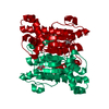
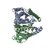
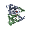
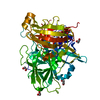
| Title | Crystal Structure of the Beta Carbonic Anhydrase psCA3 isolated from Pseudomonas aeruginosa - alternate crystal packing form | ||||||
 Components Components | Carbonic anhydrase | ||||||
 Keywords Keywords | LYASE / psCA3 / crystal packing / beta-carbonic anhydrase | ||||||
| Function / homology |  Function and homology information Function and homology informationcarbon utilization / carbonic anhydrase / carbonate dehydratase activity / zinc ion binding Similarity search - Function | ||||||
| Biological species |   Pseudomonas aeruginosa (bacteria) Pseudomonas aeruginosa (bacteria) | ||||||
| Method |  X-RAY DIFFRACTION / SYNCHROTRON / MOLECULAR REPLACEMENT / Resolution: 2.585 Å X-RAY DIFFRACTION / SYNCHROTRON / MOLECULAR REPLACEMENT / Resolution: 2.585 Å | ||||||
 Authors Authors | Pinard, M.A. / Kurian, J.J. / Aggarwal, M. / Agbandje-McKenna, M. / McKenna, R. | ||||||
| Funding support |  United States, 1items United States, 1items
| ||||||
 Citation Citation | Journal: Acta Crystallogr.,Sect.F / Year: 2016 Title: Cryoannealing-induced space-group transition of crystals of the carbonic anhydrase psCA3. Authors: Pinard, M.A. / Kurian, J.J. / Aggarwal, M. / Agbandje-McKenna, M. / McKenna, R. | ||||||
| History |
|
- Structure visualization
| Structure viewer | Molecule: MolmilJmol/JSmol |
|---|
- Downloads & links
-Download
| PDBx/mmCIF format | 5jj8.cif.gz | 182.3 KB | Display | PDBx/mmCIF format |
|---|---|---|---|---|
| PDB format | pdb5jj8.ent.gz | 145.4 KB | Display | PDB format |
| PDBx/mmJSON format | 5jj8.json.gz | Tree view | PDBx/mmJSON format | |
| Others |  Other downloads Other downloads |
-Validation report
| Arichive directory | https://data.pdbj.org/pub/pdb/validation_reports/jj/5jj8ftp://data.pdbj.org/pub/pdb/validation_reports/jj/5jj8 | HTTPS FTP |
|---|
-Related structure data
| Related structure data | 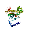 4rxyS S: Starting model for refinement |
|---|---|
| Similar structure data |
-Links
 PDBj
PDBj
- Assembly
| Deposited unit | 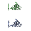
| |||||||||
|---|---|---|---|---|---|---|---|---|---|---|
| 1 | 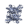
| |||||||||
| 2 | 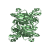
| |||||||||
| Unit cell |
| |||||||||
| Components on special symmetry positions |
|
-Components
| #1: Protein | Mass: 24250.568 Da / Num. of mol.: 2 Source method: isolated from a genetically manipulated source Source: (gene. exp.) Pseudomonas aeruginosa (bacteria)Gene: can_1, can, yadF, AO943_21535, AO946_10560, AOD73_25440, AOU28_24465, AOY09_00950, APT60_24635, ATC05_14980, AU380_02155, ERS445055_04974, HV95_21180, HV96_01220, HV97_23895, HV98_01810, HV99_ ...Gene: can_1, can, yadF, AO943_21535, AO946_10560, AOD73_25440, AOU28_24465, AOY09_00950, APT60_24635, ATC05_14980, AU380_02155, ERS445055_04974, HV95_21180, HV96_01220, HV97_23895, HV98_01810, HV99_02890, HW00_06825, HW01_20790, HW02_08065, HW03_11820, HW04_25045, HW05_22660, HW06_00905, HW07_08265, HW08_30020, HW09_16360, HW10_17975, IOMTU133_5488, PA257_5879, PA8380_53220, PAERUG_E15_London_28_01_14_01862, PAERUG_P32_London_17_VIM_2_10_11_05532, PAO1OR4768 Production host: References: UniProt: A0A072ZBL6, UniProt: Q9HVB9*PLUS, carbonic anhydrase #2: Chemical |   Mass: 65.409 Da / Num. of mol.: 2 / Source method: obtained synthetically / Formula: Zn Mass: 65.409 Da / Num. of mol.: 2 / Source method: obtained synthetically / Formula: Zn#3: Water | ChemComp-HOH / |  Mass: 18.015 Da / Num. of mol.: 22 / Source method: isolated from a natural source / Formula: H2O Mass: 18.015 Da / Num. of mol.: 22 / Source method: isolated from a natural source / Formula: H2O |
|---|
-Experimental details
-Experiment
| Experiment | Method: X-RAY DIFFRACTION / Number of used crystals: 1 |
|---|
- Sample preparation
| Crystal | Density Matthews: 2.52 Å3/Da / Density % sol: 51.11 % |
|---|---|
| Crystal grow | Temperature: 290 K / Method: vapor diffusion, sitting drop Details: Hampton Research Crystal Screen 2, PEG/Ion, PEG/Ion 2, an in-house sodium citrate screen (screening conditions varied from 1.1 to 1.8 M sodium citrate, Tris-HCl pH 7.1-8.1), and an in-house ...Details: Hampton Research Crystal Screen 2, PEG/Ion, PEG/Ion 2, an in-house sodium citrate screen (screening conditions varied from 1.1 to 1.8 M sodium citrate, Tris-HCl pH 7.1-8.1), and an in-house ammonium sulfate screen (screening conditions varied from 1.8 M to 2.8 M ammonium sulfate, 0.1 M malic acid, 0.1 M imidazole pH 7.0-8.5) prepared by the Rigaku Alchemist DT were used to screen for optimum crystallization conditions. |
-Data collection
| Diffraction | Mean temperature: 100 K |
|---|---|
| Diffraction source | Source: SYNCHROTRON / Site: CHESS / Beamline: F1 / Wavelength: 0.9177 Å |
| Detector | Type: ADSC QUANTUM 270 / Detector: CCD / Date: Mar 25, 2013 |
| Radiation | Protocol: SINGLE WAVELENGTH / Monochromatic (M) / Laue (L): M / Scattering type: x-ray |
| Radiation wavelength | Wavelength: 0.9177 Å / Relative weight: 1 |
| Reflection | Resolution: 2.585→35.553 Å / Num. obs: 14191 / % possible obs: 91.48 % / Redundancy: 3.2 % / Net I/σ(I): 17.3 |
- Processing
| Software |
| ||||||||||||||||||||||||||||||||||||||||||
|---|---|---|---|---|---|---|---|---|---|---|---|---|---|---|---|---|---|---|---|---|---|---|---|---|---|---|---|---|---|---|---|---|---|---|---|---|---|---|---|---|---|---|---|
| Refinement | Method to determine structure: MOLECULAR REPLACEMENT Starting model: 4RXY Resolution: 2.585→35.553 Å / SU ML: 0.33 / Cross valid method: FREE R-VALUE / σ(F): 1.34 / Phase error: 32.09
| ||||||||||||||||||||||||||||||||||||||||||
| Solvent computation | Shrinkage radii: 0.9 Å / VDW probe radii: 1.11 Å | ||||||||||||||||||||||||||||||||||||||||||
| Refinement step | Cycle: LAST / Resolution: 2.585→35.553 Å
| ||||||||||||||||||||||||||||||||||||||||||
| Refine LS restraints |
| ||||||||||||||||||||||||||||||||||||||||||
| LS refinement shell |
| ||||||||||||||||||||||||||||||||||||||||||
| Refinement TLS params. | Method: refined / Origin x: 16.6609 Å / Origin y: -9.0053 Å / Origin z: -16.6188 Å
| ||||||||||||||||||||||||||||||||||||||||||
| Refinement TLS group | Selection details: all |
