[English] 日本語
Yorodumi
- PDB-5fn7: Crystal structure of human CD45 extracellular region, domains d1-d2 -

+
Open data


ID or keywords:

Loading...

-
Basic information

Entry
Database: PDB / ID: 5fn7
TitleCrystal structure of human CD45 extracellular region, domains d1-d2
ComponentsRECEPTOR-TYPE TYROSINE-PROTEIN PHOSPHATASE C
KeywordsHYDROLASE / RECEPTOR PROTEIN TYROSINE PHOSPHATASE C / CD45 / PTPRC
Function / homology
Function and homology information


plasma membrane raft distribution / positive regulation of antigen receptor-mediated signaling pathway / membrane microdomain / regulation of protein tyrosine kinase activity / positive regulation of protein tyrosine phosphatase activity / positive regulation of hematopoietic stem cell migration / negative regulation of cytokine-mediated signaling pathway / alpha-beta T cell proliferation / negative regulation of protein tyrosine kinase activity / positive regulation of Fc receptor mediated stimulatory signaling pathway ...plasma membrane raft distribution / positive regulation of antigen receptor-mediated signaling pathway / membrane microdomain / regulation of protein tyrosine kinase activity / positive regulation of protein tyrosine phosphatase activity / positive regulation of hematopoietic stem cell migration / negative regulation of cytokine-mediated signaling pathway / alpha-beta T cell proliferation / negative regulation of protein tyrosine kinase activity / positive regulation of Fc receptor mediated stimulatory signaling pathway / negative regulation of cell adhesion involved in substrate-bound cell migration / regulation of interleukin-8 production / negative regulation of microglial cell activation / Other semaphorin interactions / negative regulation of T cell mediated cytotoxicity / positive regulation of humoral immune response mediated by circulating immunoglobulin / DN2 thymocyte differentiation / cell cycle phase transition / negative regulation of protein autophosphorylation / gamma-delta T cell differentiation / natural killer cell differentiation / positive regulation of gamma-delta T cell differentiation / transmembrane receptor protein tyrosine phosphatase activity / : / bleb / positive regulation of alpha-beta T cell proliferation / positive regulation of isotype switching to IgG isotypes / negative thymic T cell selection / stem cell development / heparan sulfate proteoglycan binding / positive thymic T cell selection / regulation of phagocytosis / positive regulation of extrinsic apoptotic signaling pathway / heterotypic cell-cell adhesion / bone marrow development / regulation of receptor signaling pathway via JAK-STAT / negative regulation of interleukin-2 production / ankyrin binding / leukocyte cell-cell adhesion / spectrin binding / response to aldosterone / positive regulation of stem cell proliferation / Phosphorylation of CD3 and TCR zeta chains / B cell proliferation / : / positive regulation of immunoglobulin production / T cell differentiation / positive regulation of protein kinase activity / hematopoietic progenitor cell differentiation / positive regulation of phagocytosis / dephosphorylation / positive regulation of B cell proliferation / extrinsic apoptotic signaling pathway / release of sequestered calcium ion into cytosol / positive regulation of T cell proliferation / positive regulation of interleukin-2 production / T cell activation / protein dephosphorylation / B cell differentiation / protein-tyrosine-phosphatase / secretory granule membrane / protein tyrosine phosphatase activity / response to gamma radiation / B cell receptor signaling pathway / negative regulation of protein kinase activity / cytoplasmic side of plasma membrane / negative regulation of ERK1 and ERK2 cascade / positive regulation of T cell mediated cytotoxicity / MAPK cascade / positive regulation of peptidyl-tyrosine phosphorylation / positive regulation of tumor necrosis factor production / heparin binding / T cell receptor signaling pathway / regulation of gene expression / defense response to virus / positive regulation of MAPK cascade / positive regulation of ERK1 and ERK2 cascade / cell surface receptor signaling pathway / regulation of cell cycle / membrane raft / external side of plasma membrane / signaling receptor binding / focal adhesion / Neutrophil degranulation / protein kinase binding / cell surface / extracellular exosome / membrane / plasma membrane
Similarity search - Function
Receptor-type tyrosine-protein phosphatase C / Protein tyrosine phosphatase, receptor type, N-terminal / Protein tyrosine phosphatase N terminal / Leukocyte receptor CD45 / Protein tyrosine phosphatase, catalytic domain / PTP type protein phosphatase domain profile. / Protein-tyrosine phosphatase / Tyrosine-specific protein phosphatase, PTPase domain / Protein-tyrosine phosphatase, catalytic / Protein tyrosine phosphatase, catalytic domain motif ...Receptor-type tyrosine-protein phosphatase C / Protein tyrosine phosphatase, receptor type, N-terminal / Protein tyrosine phosphatase N terminal / Leukocyte receptor CD45 / Protein tyrosine phosphatase, catalytic domain / PTP type protein phosphatase domain profile. / Protein-tyrosine phosphatase / Tyrosine-specific protein phosphatase, PTPase domain / Protein-tyrosine phosphatase, catalytic / Protein tyrosine phosphatase, catalytic domain motif / Tyrosine specific protein phosphatases active site. / Protein-tyrosine phosphatase, active site / Tyrosine-specific protein phosphatases domain / Tyrosine specific protein phosphatases domain profile. / Protein-tyrosine phosphatase-like / Fibronectin type III domain / Fibronectin type 3 domain / Fibronectin type-III domain profile. / Fibronectin type III / Fibronectin type III superfamily / Immunoglobulin-like fold
Similarity search - Domain/homology
: / Receptor-type tyrosine-protein phosphatase C
Similarity search - Component
Biological speciesHOMO SAPIENS (human)
MethodX-RAY DIFFRACTION / SYNCHROTRON / SAD / Resolution: 2.3 Å
AuthorsChang, V.T. / Fernandes, R.A. / Ganzinger, K.A. / Lee, S.F. / Siebold, C. / McColl, J. / Jonsson, P. / Palayret, M. / Harlos, K. / Coles, C.H. ...Chang, V.T. / Fernandes, R.A. / Ganzinger, K.A. / Lee, S.F. / Siebold, C. / McColl, J. / Jonsson, P. / Palayret, M. / Harlos, K. / Coles, C.H. / Jones, E.Y. / Lui, Y. / Huang, E. / Gilbert, R.J.C. / Klenerman, D. / Aricescu, A.R. / Davis, S.J.
CitationJournal: Nat.Immunol. / Year: 2016
Title: Initiation of T Cell Signaling by Cd45 Segregation at 'Close Contacts'.
Authors: Chang, V.T. / Fernandes, R.A. / Ganzinger, K.A. / Lee, S.F. / Siebold, C. / Mccoll, J. / Jonsson, P. / Palayret, M. / Harlos, K. / Coles, C.H. / Jones, E.Y. / Lui, Y. / Huang, E. / Gilbert, ...Authors: Chang, V.T. / Fernandes, R.A. / Ganzinger, K.A. / Lee, S.F. / Siebold, C. / Mccoll, J. / Jonsson, P. / Palayret, M. / Harlos, K. / Coles, C.H. / Jones, E.Y. / Lui, Y. / Huang, E. / Gilbert, R.J. / Klenerman, D. / Aricescu, A.R. / Davis, S.J.
History
DepositionNov 10, 2015Deposition site: PDBE / Processing site: PDBE
Revision 1.0Mar 23, 2016Provider: repository / Type: Initial release
Revision 1.1Mar 30, 2016Group: Database references
Revision 1.2May 18, 2016Group: Database references
Revision 1.3Apr 3, 2019Group: Data collection / Derived calculations ...Data collection / Derived calculations / Other / Source and taxonomy
Category: entity_src_gen / pdbx_database_proc ...entity_src_gen / pdbx_database_proc / pdbx_database_status / struct_conn
Item: _entity_src_gen.pdbx_host_org_cell_line / _pdbx_database_status.recvd_author_approval / _struct_conn.pdbx_leaving_atom_flag
Revision 1.4Jul 29, 2020Group: Data collection / Derived calculations ...Data collection / Derived calculations / Other / Structure summary
Category: chem_comp / entity ...chem_comp / entity / pdbx_chem_comp_identifier / pdbx_database_status / pdbx_entity_nonpoly / struct_conn / struct_site / struct_site_gen
Item: _chem_comp.name / _chem_comp.type ..._chem_comp.name / _chem_comp.type / _entity.pdbx_description / _pdbx_database_status.status_code_sf / _pdbx_entity_nonpoly.name / _struct_conn.conn_type_id / _struct_conn.id / _struct_conn.pdbx_dist_value / _struct_conn.pdbx_leaving_atom_flag / _struct_conn.pdbx_role / _struct_conn.ptnr1_auth_asym_id / _struct_conn.ptnr1_auth_comp_id / _struct_conn.ptnr1_auth_seq_id / _struct_conn.ptnr1_label_asym_id / _struct_conn.ptnr1_label_atom_id / _struct_conn.ptnr1_label_comp_id / _struct_conn.ptnr1_label_seq_id / _struct_conn.ptnr2_auth_asym_id / _struct_conn.ptnr2_auth_comp_id / _struct_conn.ptnr2_auth_seq_id / _struct_conn.ptnr2_label_asym_id / _struct_conn.ptnr2_label_atom_id / _struct_conn.ptnr2_label_comp_id / _struct_conn.ptnr2_label_seq_id
Description: Carbohydrate remediation / Provider: repository / Type: Remediation

-
Structure visualization

Structure viewerMolecule:
MolmilJmol/JSmol

Downloads & links

-
Assembly

Deposited unit
A: RECEPTOR-TYPE TYROSINE-PROTEIN PHOSPHATASE C
B: RECEPTOR-TYPE TYROSINE-PROTEIN PHOSPHATASE C
hetero molecules


Theoretical massNumber of molelcules
Total (without water)44,98813
Polymers42,5952
Non-polymers2,39211
Water1,892105
1
A: RECEPTOR-TYPE TYROSINE-PROTEIN PHOSPHATASE C
hetero molecules


Theoretical massNumber of molelcules
Total (without water)22,5847
Polymers21,2981
Non-polymers1,2866
Water181
TypeNameSymmetry operationNumber
identity operation1_555x,y,z1
MethodPISA
2
B: RECEPTOR-TYPE TYROSINE-PROTEIN PHOSPHATASE C
hetero molecules


Theoretical massNumber of molelcules
Total (without water)22,4046
Polymers21,2981
Non-polymers1,1065
Water181
TypeNameSymmetry operationNumber
identity operation1_555x,y,z1
MethodPISA
Unit cell
Length a, b, c (Å)149.610, 149.610, 52.730
Angle α, β, γ (deg.)90.00, 90.00, 120.00
Int Tables number173
Space group name H-MP63

-
Components

#1: Protein RECEPTOR-TYPE TYROSINE-PROTEIN PHOSPHATASE C / LEUKOCYTE COMMON ANTIGEN / L-CA / T200 / CD45


Mass: 21297.725 Da / Num. of mol.: 2 / Fragment: DOMAINS D1-D2, RESIDUES 223-392
Source method: isolated from a genetically manipulated source
Source: (gene. exp.) HOMO SAPIENS (human) / Cell: T-CELLS / Plasmid: PHLSEC / Cell line (production host): HEK293T / Production host: HOMO SAPIENS (human) / References: UniProt: P08575, protein-tyrosine-phosphatase
#2: Sugar
ChemComp-NAG / 2-acetamido-2-deoxy-beta-D-glucopyranose / N-Acetylglucosamine


Type: D-saccharide, beta linking / Mass: 221.208 Da / Num. of mol.: 9
Source method: isolated from a genetically manipulated source
Formula: C8H15NO6
IdentifierTypeProgram
DGlcpNAcbCONDENSED IUPAC CARBOHYDRATE SYMBOLGMML 1.0
N-acetyl-b-D-glucopyranosamineCOMMON NAMEGMML 1.0
b-D-GlcpNAcIUPAC CARBOHYDRATE SYMBOLPDB-CARE 1.0
GlcNAcSNFG CARBOHYDRATE SYMBOLGMML 1.0
#3: Chemical ChemComp-HG / MERCURY (II) ION / Mercury (element)


Mass: 200.590 Da / Num. of mol.: 2 / Source method: obtained synthetically / Formula: Hg
#4: Water ChemComp-HOH / water / Water


Mass: 18.015 Da / Num. of mol.: 105 / Source method: isolated from a natural source / Formula: H2O
Sequence detailsETGIEGR N-TERMINAL RESIDUES DERIVED FROM THE EXPRESSION VECTOR. GTKHHHHHH C-TERMINAL RESIDUES ...ETGIEGR N-TERMINAL RESIDUES DERIVED FROM THE EXPRESSION VECTOR. GTKHHHHHH C-TERMINAL RESIDUES DERIVED FROM THE EXPRESSION VECTOR.

-
Experimental details

-
Experiment

Experiment
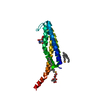
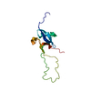
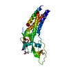
Method: X-RAY DIFFRACTION / Number of used crystals: 1

-
Sample preparation

CrystalDensity Matthews: 4.26 Å3/Da / Density % sol: 71 % / Description: NONE
Crystal growpH: 8
Details: 0.1M TRIS, PH=8, 2.4M (NH4)2SO4, 6% ETHYLENE GLYCOL

-
Data collection

DiffractionMean temperature: 100 K
Diffraction sourceSource: SYNCHROTRON / Site: ESRF / Beamline: ID23-1 / Wavelength: 0.9497
DetectorType: ADSC CCD / Detector: CCD / Date: Feb 11, 2008
RadiationProtocol: SINGLE WAVELENGTH / Monochromatic (M) / Laue (L): M / Scattering type: x-ray
Radiation wavelengthWavelength: 0.9497 Å / Relative weight: 1
ReflectionResolution: 2.3→43 Å / Num. obs: 30027 / % possible obs: 99.2 % / Observed criterion σ(I): 2.6 / Redundancy: 9.2 % / Biso Wilson estimate: 50.89 Å2 / Rmerge(I) obs: 0.08 / Net I/σ(I): 17.6
Reflection shellResolution: 2.3→2.36 Å / Redundancy: 9.1 % / Rmerge(I) obs: 0.92 / Mean I/σ(I) obs: 2.6 / % possible all: 99.8

-
Processing

Software
NameVersionClassification
HKL-2000data reduction
HKL-2000data scaling
SHELXDphasing
autoSHARPphasing
BUSTER2.11.2refinement
RefinementMethod to determine structure: SAD
Starting model: NONE

Resolution: 2.3→43 Å / Cor.coef. Fo:Fc: 0.9241 / Cor.coef. Fo:Fc free: 0.9112 / SU R Cruickshank DPI: 0.188 / Cross valid method: THROUGHOUT / σ(F): 0 / SU R Blow DPI: 0.187 / SU Rfree Blow DPI: 0.171 / SU Rfree Cruickshank DPI: 0.173
Details: IDEAL-DIST CONTACT TERM CONTACT SETUP. RESIDUE TYPES WITHOUT CCP4 ATOM TYPE IN LIBRARY=HG. NUMBER OF ATOMS WITH PROPER CCP4 ATOM TYPE=2975. NUMBER WITH APPROX DEFAUL CCP4 ATOM TYPE=0. NUMBER ...Details: IDEAL-DIST CONTACT TERM CONTACT SETUP. RESIDUE TYPES WITHOUT CCP4 ATOM TYPE IN LIBRARY=HG. NUMBER OF ATOMS WITH PROPER CCP4 ATOM TYPE=2975. NUMBER WITH APPROX DEFAUL CCP4 ATOM TYPE=0. NUMBER TREATED BY BAD NON-BONDED CONTACTS=2.
RfactorNum. reflection% reflectionSelection details
Rfree0.2385 1524 5.08 %RANDOM
Rwork0.2015 ---
obs0.2033 30020 99.17 %-
Displacement parametersBiso mean: 58.89 Å2
Baniso -1Baniso -2Baniso -3
1--7.3634 Å20 Å20 Å2
2---7.3634 Å20 Å2
3---14.7268 Å2
Refine analyzeLuzzati coordinate error obs: 0.401 Å
Refinement stepCycle: LAST / Resolution: 2.3→43 Å
ProteinNucleic acidLigandSolventTotal
Num. atoms2744 0 128 105 2977
Refine LS restraints
Refine-IDTypeDev idealNumberRestraint functionWeight
X-RAY DIFFRACTIONt_bond_d0.012941HARMONIC2
X-RAY DIFFRACTIONt_angle_deg1.324007HARMONIC2
X-RAY DIFFRACTIONt_dihedral_angle_d1078SINUSOIDAL2
X-RAY DIFFRACTIONt_incorr_chiral_ct
X-RAY DIFFRACTIONt_pseud_angle
X-RAY DIFFRACTIONt_trig_c_planes100HARMONIC2
X-RAY DIFFRACTIONt_gen_planes410HARMONIC5
X-RAY DIFFRACTIONt_it2941HARMONIC20
X-RAY DIFFRACTIONt_nbd1SEMIHARMONIC5
X-RAY DIFFRACTIONt_omega_torsion3.48
X-RAY DIFFRACTIONt_other_torsion18.05
X-RAY DIFFRACTIONt_improper_torsion
X-RAY DIFFRACTIONt_chiral_improper_torsion439SEMIHARMONIC5
X-RAY DIFFRACTIONt_sum_occupancies
X-RAY DIFFRACTIONt_utility_distance
X-RAY DIFFRACTIONt_utility_angle
X-RAY DIFFRACTIONt_utility_torsion
X-RAY DIFFRACTIONt_ideal_dist_contact3220SEMIHARMONIC4
LS refinement shellResolution: 2.3→2.38 Å / Total num. of bins used: 15
RfactorNum. reflection% reflection
Rfree0.2738 145 4.95 %
Rwork0.2463 2782 -
all0.2477 2927 -
obs--99.17 %
Refinement TLS params.

Method: refined / Refine-ID: X-RAY DIFFRACTION

IDL112)L122)L132)L222)L232)L332)S11 (Å °)S12 (Å °)S13 (Å °)S21 (Å °)S22 (Å °)S23 (Å °)S31 (Å °)S32 (Å °)S33 (Å °)T112)T122)T132)T222)T232)T332)Origin x (Å)Origin y (Å)Origin z (Å)
11.17720.11061.25640.61641.31132.71620.0686-0.08060.11330.1108-0.1173-0.01260.24830.18310.0487-0.0299-0.19320.0217-0.07260.0558-0.09727.3305170.0242-22.4687
20.6668-0.33180.34741.84942.20344.2974-0.05510.0208-0.0807-0.19250.0120.08990.07170.39390.04310.0224-0.08630.0153-0.23860.0608-0.120814.9541147.0396-60.1529
Refinement TLS group
IDRefine-IDRefine TLS-IDSelection details
1X-RAY DIFFRACTION1{ A|2 - A|173 }
2X-RAY DIFFRACTION2{ B|2 - B|172 }

+
About Yorodumi

-
News

-
Feb 9, 2022. New format data for meta-information of EMDB entries

New format data for meta-information of EMDB entries

  • Version 3 of the EMDB header file is now the official format.
  • The previous official version 1.9 will be removed from the archive.

Related info.:EMDB header

External links:wwPDB to switch to version 3 of the EMDB data model

-
Aug 12, 2020. Covid-19 info

Covid-19 info

URL: https://pdbj.org/emnavi/covid19.php

New page: Covid-19 featured information page in EM Navigator.

Related info.:Covid-19 info / Mar 5, 2020. Novel coronavirus structure data

+
Mar 5, 2020. Novel coronavirus structure data

Novel coronavirus structure data

Related info.:Yorodumi Speices / Aug 12, 2020. Covid-19 info

External links:COVID-19 featured content - PDBj / Molecule of the Month (242):Coronavirus Proteases

+
Jan 31, 2019. EMDB accession codes are about to change! (news from PDBe EMDB page)

EMDB accession codes are about to change! (news from PDBe EMDB page)

  • The allocation of 4 digits for EMDB accession codes will soon come to an end. Whilst these codes will remain in use, new EMDB accession codes will include an additional digit and will expand incrementally as the available range of codes is exhausted. The current 4-digit format prefixed with “EMD-” (i.e. EMD-XXXX) will advance to a 5-digit format (i.e. EMD-XXXXX), and so on. It is currently estimated that the 4-digit codes will be depleted around Spring 2019, at which point the 5-digit format will come into force.
  • The EM Navigator/Yorodumi systems omit the EMD- prefix.

Related info.:Q: What is EMD? / ID/Accession-code notation in Yorodumi/EM Navigator

External links:EMDB Accession Codes are Changing Soon! / Contact to PDBj

+
Jul 12, 2017. Major update of PDB

Major update of PDB

  • wwPDB released updated PDB data conforming to the new PDBx/mmCIF dictionary.
  • This is a major update changing the version number from 4 to 5, and with Remediation, in which all the entries are updated.
  • In this update, many items about electron microscopy experimental information are reorganized (e.g. em_software).
  • Now, EM Navigator and Yorodumi are based on the updated data.

External links:wwPDB Remediation / Enriched Model Files Conforming to OneDep Data Standards Now Available in the PDB FTP Archive

-
Yorodumi

Thousand views of thousand structures

  • Yorodumi is a browser for structure data from EMDB, PDB, SASBDB, etc.
  • This page is also the successor to EM Navigator detail page, and also detail information page/front-end page for Omokage search.
  • The word "yorodu" (or yorozu) is an old Japanese word meaning "ten thousand". "mi" (miru) is to see.

Related info.:EMDB / PDB / SASBDB / Comparison of 3 databanks / Yorodumi Search / Aug 31, 2016. New EM Navigator & Yorodumi / Yorodumi Papers / Jmol/JSmol / Function and homology information / Changes in new EM Navigator and Yorodumi

Read more