 Movie
Movie Controller


+ Open data

- Basic information
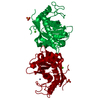
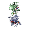
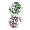
| Entry | Database: PDB / ID: 5e8c | ||||||
|---|---|---|---|---|---|---|---|
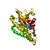
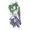
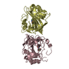
| Title | pseudorabies virus nuclear egress complex, pUL31, pUL34 | ||||||
 Components Components |
| ||||||
 Keywords Keywords |  VIRAL PROTEIN / herpesviruses / pseudorabies virus / PrV / nuclear egress / curvature / membrane remodelling / NEC / zinc finger motif / pUL31 / pUL34 / vesicle formation / trasncription VIRAL PROTEIN / herpesviruses / pseudorabies virus / PrV / nuclear egress / curvature / membrane remodelling / NEC / zinc finger motif / pUL31 / pUL34 / vesicle formation / trasncription | ||||||
| Function / homology |  Function and homology information Function and homology informationhost cell nuclear inner membrane / viral budding from nuclear membrane / membrane / metal ion bindingSimilarity search - Function | ||||||
| Biological species |   Suid herpesvirus 1 Suid herpesvirus 1 | ||||||
| Method | X-RAY DIFFRACTION / SYNCHROTRON / SAD / Resolution: 2.9 Å | ||||||
 Authors Authors | Zeev-Ben-Mordehai, T. / Cheleski, J. / Whittle, C. / El Omari, K. / Harlos, K. / Hagen, C. / Klupp, B. / Mettenleiter, T.C. / Gruenewald, K. | ||||||
| Funding support |  United Kingdom, 1items United Kingdom, 1items
| ||||||
 Citation Citation | Journal: Cell Rep / Year: 2015 Title: Crystal Structure of the Herpesvirus Nuclear Egress Complex Provides Insights into Inner Nuclear Membrane Remodeling. Authors: Tzviya Zeev-Ben-Mordehai / Marion Weberruß / Michael Lorenz / Juliana Cheleski / Teresa Hellberg / Cathy Whittle / Kamel El Omari / Daven Vasishtan / Kyle C Dent / Karl Harlos / Kati ...Authors: Tzviya Zeev-Ben-Mordehai / Marion Weberruß / Michael Lorenz / Juliana Cheleski / Teresa Hellberg / Cathy Whittle / Kamel El Omari / Daven Vasishtan / Kyle C Dent / Karl Harlos / Kati Franzke / Christoph Hagen / Barbara G Klupp / Wolfram Antonin / Thomas C Mettenleiter / Kay Grünewald /  Abstract: Although nucleo-cytoplasmic transport is typically mediated through nuclear pore complexes, herpesvirus capsids exit the nucleus via a unique vesicular pathway. Together, the conserved herpesvirus ...Although nucleo-cytoplasmic transport is typically mediated through nuclear pore complexes, herpesvirus capsids exit the nucleus via a unique vesicular pathway. Together, the conserved herpesvirus proteins pUL31 and pUL34 form the heterodimeric nuclear egress complex (NEC), which, in turn, mediates the formation of tight-fitting membrane vesicles around capsids at the inner nuclear membrane. Here, we present the crystal structure of the pseudorabies virus NEC. The structure revealed that a zinc finger motif in pUL31 and an extensive interaction network between the two proteins stabilize the complex. Comprehensive mutational analyses, characterized both in situ and in vitro, indicated that the interaction network is not redundant but rather complementary. Fitting of the NEC crystal structure into the recently determined cryoEM-derived hexagonal lattice, formed in situ by pUL31 and pUL34, provided details on the molecular basis of NEC coat formation and inner nuclear membrane remodeling. | ||||||
| History |
|
- Structure visualization
| Structure viewer | Molecule: MolmilJmol/JSmol |
|---|
- Downloads & links
-Download
| PDBx/mmCIF format | 5e8c.cif.gz | 171.7 KB | Display | PDBx/mmCIF format |
|---|---|---|---|---|
| PDB format | pdb5e8c.ent.gz | 142.3 KB | Display | PDB format |
| PDBx/mmJSON format | 5e8c.json.gz | Tree view | PDBx/mmJSON format | |
| Others |  Other downloads Other downloads |
-Validation report
| Arichive directory | https://data.pdbj.org/pub/pdb/validation_reports/e8/5e8cftp://data.pdbj.org/pub/pdb/validation_reports/e8/5e8c | HTTPS FTP |
|---|
-Related structure data


-Links
 PDBj
PDBj- Assembly
| Deposited unit | 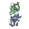
| ||||||||
|---|---|---|---|---|---|---|---|---|---|
| 1 |
| ||||||||
| Unit cell |
|
-Components
| #1: Protein | Mass: 27668.900 Da / Num. of mol.: 1 Source method: isolated from a genetically manipulated source Source: (gene. exp.) Suid herpesvirus 1 / Gene: UL31 / Production host:  Escherichia coli (E. coli) / References: UniProt: G3G955 Escherichia coli (E. coli) / References: UniProt: G3G955 |
|---|---|
| #2: Protein | Mass: 19378.076 Da / Num. of mol.: 1 Source method: isolated from a genetically manipulated source Source: (gene. exp.) Suid herpesvirus 1 / Gene: UL34 / Production host: Escherichia coli (E. coli) / References: UniProt: G3G8R3 |
| #3: Chemical | ChemComp-ZN /   Mass: 65.409 Da / Num. of mol.: 1 / Source method: obtained synthetically / Formula: Zn Mass: 65.409 Da / Num. of mol.: 1 / Source method: obtained synthetically / Formula: Zn |
| #4: Chemical | ChemComp-CL / Chloride  Mass: 35.453 Da / Num. of mol.: 1 / Source method: obtained synthetically / Formula: Cl Mass: 35.453 Da / Num. of mol.: 1 / Source method: obtained synthetically / Formula: Cl |
-Experimental details
-Experiment
| Experiment | Method: X-RAY DIFFRACTION / Number of used crystals: 1 |
|---|
- Sample preparation
| Crystal | Density Matthews: 2.42 Å3/Da / Density % sol: 49.14 % |
|---|---|
| Crystal grow | Temperature: 298 K / Method: vapor diffusion, sitting drop Details: 16% PEGG6000, MMT buffer system pH7.0, 1M Lithium Chloride |
-Data collection
| Diffraction | Mean temperature: 80 K |
|---|---|
| Diffraction source | Source: SYNCHROTRON / Site: Diamond / Beamline: I04 / Wavelength: 0.97902 Å |
| Detector | Type: PSI PILATUS 6M / Detector: PIXEL / Date: Feb 18, 2015 |
| Radiation | Protocol: SINGLE WAVELENGTH / Monochromatic (M) / Laue (L): M / Scattering type: x-ray |
| Radiation wavelength | Wavelength: 0.97902 Å / Relative weight: 1 |
| Reflection | Resolution: 2.9→70 Å / Num. obs: 10771 / % possible obs: 99.9 % / Redundancy: 227 % / Biso Wilson estimate: 87 Å2 / Rmerge(I) obs: 0.308 / Net I/σ(I): 35.6 |
| Reflection shell | Resolution: 2.9→2.98 Å / Redundancy: 209.1 % / Rmerge(I) obs: 3.46 / Mean I/σ(I) obs: 4.2 / Num. unique all: 777 / % possible all: 99.9 |
- Processing
| Software |
| ||||||||||||||||||||||||||||||||||||||||||||||||||||||||||||||||||||||||||||||||||||||||||||||||||||||||||||
|---|---|---|---|---|---|---|---|---|---|---|---|---|---|---|---|---|---|---|---|---|---|---|---|---|---|---|---|---|---|---|---|---|---|---|---|---|---|---|---|---|---|---|---|---|---|---|---|---|---|---|---|---|---|---|---|---|---|---|---|---|---|---|---|---|---|---|---|---|---|---|---|---|---|---|---|---|---|---|---|---|---|---|---|---|---|---|---|---|---|---|---|---|---|---|---|---|---|---|---|---|---|---|---|---|---|---|---|---|---|
| Refinement | Method to determine structure: SAD / Resolution: 2.9→69.97 Å / Cor.coef. Fo:Fc: 0.9081 / Cor.coef. Fo:Fc free: 0.8809 / Cross valid method: THROUGHOUT / σ(F): 0 / SU Rfree Blow DPI: 0.413
| ||||||||||||||||||||||||||||||||||||||||||||||||||||||||||||||||||||||||||||||||||||||||||||||||||||||||||||
| Displacement parameters | Biso max: 205.3 Å2 / Biso mean: 92.7 Å2 / Biso min: 49.92 Å2
| ||||||||||||||||||||||||||||||||||||||||||||||||||||||||||||||||||||||||||||||||||||||||||||||||||||||||||||
| Refine analyze | Luzzati coordinate error obs: 0.419 Å | ||||||||||||||||||||||||||||||||||||||||||||||||||||||||||||||||||||||||||||||||||||||||||||||||||||||||||||
| Refinement step | Cycle: final / Resolution: 2.9→69.97 Å
| ||||||||||||||||||||||||||||||||||||||||||||||||||||||||||||||||||||||||||||||||||||||||||||||||||||||||||||
| Refine LS restraints |
| ||||||||||||||||||||||||||||||||||||||||||||||||||||||||||||||||||||||||||||||||||||||||||||||||||||||||||||
| LS refinement shell | Resolution: 2.9→3.24 Å / Total num. of bins used: 5
| ||||||||||||||||||||||||||||||||||||||||||||||||||||||||||||||||||||||||||||||||||||||||||||||||||||||||||||
| Refinement TLS params. | Method: refined / Refine-ID: X-RAY DIFFRACTION
| ||||||||||||||||||||||||||||||||||||||||||||||||||||||||||||||||||||||||||||||||||||||||||||||||||||||||||||
| Refinement TLS group |
|
