- PDB-5cqe: 2.1 Angstrom resolution crystal structure of matrix protein 1 (M1... -
+
Open data
ID or keywords:
Loading...
-
Basic information
Entry
Database: PDB / ID: 5cqe
Title
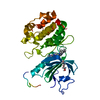
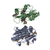
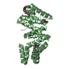
2.1 Angstrom resolution crystal structure of matrix protein 1 (M1; residues 1-164) from Influenza A virus (A/Puerto Rico/8/34(H1N1))
Components
Matrix protein 1
Keywords
VIRAL PROTEIN / matrix protein 1 / Influenza A virus (A/Puerto Rico/8/34(H1N1)) / CSGID / virulence / pathogenesis / Structural Genomics / Center for Structural Genomics of Infectious Diseases
Function / homology
Function and homology information
Assembly of Viral Components at the Budding Site / Influenza Infection / Fusion of the Influenza Virion to the Host Cell Endosome / Release / Budding / Packaging of Eight RNA Segments / Uncoating of the Influenza Virion / Entry of Influenza Virion into Host Cell via Endocytosis / Viral RNP Complexes in the Host Cell Nucleus / NEP/NS2 Interacts with the Cellular Export Machinery ...Assembly of Viral Components at the Budding Site / Influenza Infection / Fusion of the Influenza Virion to the Host Cell Endosome / Release / Budding / Packaging of Eight RNA Segments / Uncoating of the Influenza Virion / Entry of Influenza Virion into Host Cell via Endocytosis / Viral RNP Complexes in the Host Cell Nucleus / NEP/NS2 Interacts with the Cellular Export Machinery / Viral mRNA Translation / viral budding from plasma membrane / structural constituent of virion / host cell nucleus / virion membrane / RNA binding / extracellular region / plasma membrane Similarity search - Function
Resolution: 2.1→29.47 Å / Cor.coef. Fo:Fc: 0.967 / Cor.coef. Fo:Fc free: 0.962 / SU B: 8.136 / SU ML: 0.104 / Cross valid method: THROUGHOUT / ESU R: 0.184 / ESU R Free: 0.148 / Stereochemistry target values: MAXIMUM LIKELIHOOD / Details: HYDROGENS HAVE BEEN ADDED IN THE RIDING POSITIONS
Rfactor
Num. reflection
% reflection
Selection details
Rfree
0.19745
1086
4.8 %
RANDOM
Rwork
0.17491
-
-
-
obs
0.17603
21413
99.88 %
-
Solvent computation
Ion probe radii: 0.8 Å / Shrinkage radii: 0.8 Å / VDW probe radii: 1.2 Å / Solvent model: BABINET MODEL WITH MASK
 Movie
Movie Controller

 Yorodumi
 Open data
 Basic information
 Components
 Keywords
 VIRAL PROTEIN / matrix protein 1 / Influenza A virus (A/Puerto Rico/8/34(H1N1)) / CSGID /
  Function and homology information

 Authors
 Citation
 Structure visualization
 Downloads & links
 Other downloads


 PDBj
PDBj


 Assembly


 Mass: 35.453 Da / Num. of mol.: 3 / Source method: obtained synthetically / Formula: Cl
 Mass: 122.143 Da / Num. of mol.: 1 / Source method: obtained synthetically / Formula: C4H12NO3 / Comment: pH buffer*YM
 Mass: 62.068 Da / Num. of mol.: 1 / Source method: obtained synthetically / Formula: C2H6O2
 Mass: 106.120 Da / Num. of mol.: 1 / Source method: obtained synthetically / Formula: C4H10O3
 Sample preparation
 / Beamline: 21-ID-G / Wavelength: 0.97856 Å
 Processing