- PDB-4zwr: Crystal Structure of the Bacteriophage T4 recombination mediator ... -
+
Open data
ID or keywords:
Loading...
-
Basic information
Entry
Database: PDB / ID: 4zwr
Title
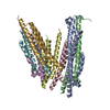
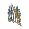
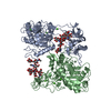
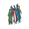
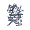
Crystal Structure of the Bacteriophage T4 recombination mediator protein UvsY, Lattice Type II
Components
Recombination protein uvsYGenetic recombination
Keywords
VIRAL PROTEIN / recombination / DNA binding / homo-heptamer / asymmetry / alpha barrel
Function / homology
Recombination, repair and ssDNA binding protein UvsY / Recombination, repair and ssDNA binding protein UvsY / DNA biosynthetic process / DNA recombination / DNA binding / Recombination protein uvsY
Function and homology information
Biological species
Enterobacteria phage T4 (virus)
Method
X-RAY DIFFRACTION / SYNCHROTRON / SAD / Resolution: 3.3857 Å
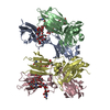
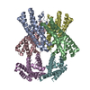
A: Recombination protein uvsY B: Recombination protein uvsY C: Recombination protein uvsY D: Recombination protein uvsY E: Recombination protein uvsY F: Recombination protein uvsY G: Recombination protein uvsY
Mass: 18364.889 Da / Num. of mol.: 7 Source method: isolated from a genetically manipulated source Source: (gene. exp.) Enterobacteria phage T4 (virus) / Gene: uvsY / Plasmid: pET28 / Production host: Escherichia coli BL21(DE3) (bacteria) / References: UniProt: P04537
-
Experimental details
-
Experiment
Experiment
Method: X-RAY DIFFRACTION
-
Sample preparation
Crystal
Density Matthews: 3.13 Å3/Da / Density % sol: 60.68 %
Crystal grow
Temperature: 277 K / Method: vapor diffusion, sitting drop / pH: 6.4 Details: 1:1 Se-Met-labeled UvsY (12 mg/mL in 0.5 M sodium chloride, 15 mM HEPES, pH 8.2, 2 mM DTT) to 18% MPD, 225 mM sodium chloride, 0.1 M MES, pH 6.4, sample subjected to reductive alkylation ...Details: 1:1 Se-Met-labeled UvsY (12 mg/mL in 0.5 M sodium chloride, 15 mM HEPES, pH 8.2, 2 mM DTT) to 18% MPD, 225 mM sodium chloride, 0.1 M MES, pH 6.4, sample subjected to reductive alkylation with formaldehyde prior to crystallization
-
Data collection
Diffraction
Mean temperature: 100 K
Diffraction source
Source: SYNCHROTRON / Site: ALS / Beamline: 8.2.2 / Wavelength: 0.9793 Å
Method to determine structure: SAD / Resolution: 3.3857→19.911 Å / SU ML: 0.41 / Cross valid method: FREE R-VALUE / σ(F): 1.35 / Phase error: 29.77 / Stereochemistry target values: ML Details: THE X-RAY DIFFRACTION DATA EXHIBITED STRONG ANISOTROPY. REFINEMENT WAS PERFORMED AGAINST ELLIPTICALLY TRUNCATED DATA (RESOLUTION LIMITS OF 1/5.0, 1/3.8, AND 1/3.4 A IN A*, B*, C*, ...Details: THE X-RAY DIFFRACTION DATA EXHIBITED STRONG ANISOTROPY. REFINEMENT WAS PERFORMED AGAINST ELLIPTICALLY TRUNCATED DATA (RESOLUTION LIMITS OF 1/5.0, 1/3.8, AND 1/3.4 A IN A*, B*, C*, RESPECTIVELY) CORRECTED BY ANISOTROPIC SCALE FACTORS AND AN ISOTROPIC B OF -52.54 A2 USING THE UCLA DIFFRACTION ANISOTROPY SERVER (STRONG ET AL., 2006).
Rfactor
Num. reflection
% reflection
Rfree
0.2665
698
5 %
Rwork
0.2391
13260
-
obs
0.2405
13958
61.31 %
Solvent computation
Shrinkage radii: 0.9 Å / VDW probe radii: 1.11 Å / Solvent model: FLAT BULK SOLVENT MODEL
 Movie
Movie Controller

 Yorodumi
 Open data
 Basic information
 Components
 Genetic recombination
  Keywords
 Function and homology information
 Authors
 United States, 3items
  Citation
 Structure visualization
 Downloads & links
 Other downloads
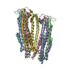


 PDBj
PDBj Assembly
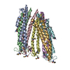

 Sample preparation
 Processing