 Movie
Movie Controller

[English] 日本語
 Yorodumi
- PDB-4wpk: Crystal structure of Mycobacterium tuberculosis uracil-DNA glycos... -
+ Open data

- Basic information
| Entry | Database: PDB / ID: 4wpk | ||||||
|---|---|---|---|---|---|---|---|
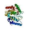
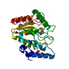
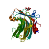
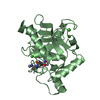
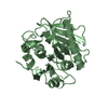
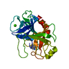
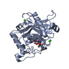
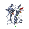
| Title | Crystal structure of Mycobacterium tuberculosis uracil-DNA glycosylase, Form I | ||||||
 Components Components | Uracil-DNA glycosylase | ||||||
 Keywords Keywords | HYDROLASE / DNA-repair / Excision repair / Conformational selection / Ligand-binding | ||||||
| Function / homology |  Function and homology information Function and homology informationbase-excision repair, AP site formation via deaminated base removal / uracil-DNA glycosylase / uracil DNA N-glycosylase activity / base-excision repair / cytoplasm Similarity search - Function | ||||||
| Biological species |  Mycobacterium tuberculosis H37Rv (bacteria) Mycobacterium tuberculosis H37Rv (bacteria) | ||||||
| Method |  X-RAY DIFFRACTION / SYNCHROTRON / Resolution: 0.98 Å X-RAY DIFFRACTION / SYNCHROTRON / Resolution: 0.98 Å | ||||||
 Authors Authors | Arif, S.M. / Geethanandan, K. / Mishra, P. / Surolia, A. / Varshney, U. / Vijayan, M. | ||||||
 Citation Citation | Journal: Acta Crystallogr.,Sect.D / Year: 2015 Title: Structural plasticity in Mycobacterium tuberculosis uracil-DNA glycosylase (MtUng) and its functional implications. Authors: Arif, S.M. / Geethanandan, K. / Mishra, P. / Surolia, A. / Varshney, U. / Vijayan, M. #1: Journal: Acta Crystallogr.,Sect.F / Year: 2010Title: Structure of uracil-DNA glycosylase from Mycobacterium tuberculosis: insights into interactions with ligands. Authors: Kaushal, P.S. / Talawar, R.K. / Varshney, U. / Vijayan, M. #2: Journal: Acta Crystallogr.,Sect.D / Year: 2008Title: Unique features of the structure and interactions of mycobacterial uracil-DNA glycosylase: structure of a complex of the Mycobacterium tuberculosis enzyme in comparison with those from other sources. Authors: Kaushal, P.S. / Talawar, R.K. / Krishna, P.D. / Varshney, U. / Vijayan, M. #3: Journal: Acta Crystallogr.,Sect.D / Year: 2002Title: Domain closure and action of uracil DNA glycosylase (UDG): structures of new crystal forms containing the Escherichia coli enzyme and a comparative study of the known structures involving UDG. Authors: Saikrishnan, K. / Bidya Sagar, M. / Ravishankar, R. / Roy, S. / Purnapatre, K. / Handa, P. / Varshney, U. / Vijayan, M. #4: Journal: Nucleic Acids Res. / Year: 1998Title: X-ray analysis of a complex of Escherichia coli uracil DNA glycosylase (EcUDG) with a proteinaceous inhibitor. The structure elucidation of a prokaryotic UDG. Authors: Ravishankar, R. / Bidya Sagar, M. / Roy, S. / Purnapatre, K. / Handa, P. / Varshney, U. / Vijayan, M. | ||||||
| History |
|
- Structure visualization
| Structure viewer | Molecule: MolmilJmol/JSmol |
|---|
- Downloads & links
-Download
| PDBx/mmCIF format | 4wpk.cif.gz | 121.7 KB | Display | PDBx/mmCIF format |
|---|---|---|---|---|
| PDB format | pdb4wpk.ent.gz | 92.7 KB | Display | PDB format |
| PDBx/mmJSON format | 4wpk.json.gz | Tree view | PDBx/mmJSON format | |
| Others |  Other downloads Other downloads |
-Validation report
| Arichive directory | https://data.pdbj.org/pub/pdb/validation_reports/wp/4wpkftp://data.pdbj.org/pub/pdb/validation_reports/wp/4wpk | HTTPS FTP |
|---|
-Related structure data
| Related structure data | 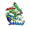 4wplC 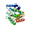 4wruC 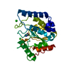 4wrvC 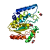 4wrwC 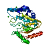 4wrxC 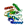 4wryC 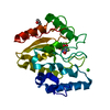 4wrzC  4ws0C  4ws1C 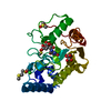 4ws2C 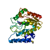 4ws3C 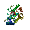 4ws4C 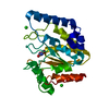 4ws5C 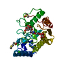 4ws6C 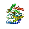 4ws7C 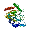 4ws8C 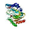 3a7nS S: Starting model for refinement C: citing same article ( |
|---|---|
| Similar structure data |

-Links
 PDBj
PDBj- Assembly
| Deposited unit | 
| ||||||||
|---|---|---|---|---|---|---|---|---|---|
| 1 |
| ||||||||
| Unit cell |
|
-Components
| #1: Protein | Mass: 25813.543 Da / Num. of mol.: 1 Source method: isolated from a genetically manipulated source Source: (gene. exp.) Mycobacterium tuberculosis H37Rv (bacteria)Gene: ung, Rv2976c, MTCY349.11 / Plasmid: pRSETB MTUUDG / Production host: |
|---|---|
| #2: Chemical | ChemComp-CIT /   Mass: 192.124 Da / Num. of mol.: 1 / Source method: obtained synthetically / Formula: C6H8O7 Mass: 192.124 Da / Num. of mol.: 1 / Source method: obtained synthetically / Formula: C6H8O7 |
| #3: Chemical | ChemComp-NA /   Mass: 22.990 Da / Num. of mol.: 1 / Source method: obtained synthetically / Formula: Na Mass: 22.990 Da / Num. of mol.: 1 / Source method: obtained synthetically / Formula: Na |
| #4: Water | ChemComp-HOH /  Mass: 18.015 Da / Num. of mol.: 347 / Source method: isolated from a natural source / Formula: H2O Mass: 18.015 Da / Num. of mol.: 347 / Source method: isolated from a natural source / Formula: H2O |
-Experimental details
-Experiment
| Experiment | Method: X-RAY DIFFRACTION |
|---|
- Sample preparation
| Crystal | Density Matthews: 2.09 Å3/Da / Density % sol: 41.24 % |
|---|---|
| Crystal grow | Temperature: 293 K / Method: microbatch / pH: 6.5 / Details: 1.6 M Sodium citrate tribasic dihydrate |
-Data collection
| Diffraction | Mean temperature: 100 K |
|---|---|
| Diffraction source | Source: SYNCHROTRON / Site: ESRF  / Beamline: BM14 / Wavelength: 0.72932 Å / Beamline: BM14 / Wavelength: 0.72932 Å |
| Detector | Type: MARMOSAIC 225 mm CCD / Detector: CCD / Date: Apr 25, 2013 |
| Radiation | Protocol: SINGLE WAVELENGTH / Monochromatic (M) / Laue (L): M / Scattering type: x-ray |
| Radiation wavelength | Wavelength: 0.72932 Å / Relative weight: 1 |
| Reflection | Resolution: 0.98→23.74 Å / Num. obs: 115505 / % possible obs: 100 % / Redundancy: 4.9 % / Net I/σ(I): 9.4 |
| Reflection shell | Resolution: 0.98→1.03 Å / Redundancy: 4.5 % / Mean I/σ(I) obs: 2.4 / % possible all: 100 |
- Processing
| Software | Name: REFMAC / Version: 5.8.0049 / Classification: refinement | ||||||||||||||||||||||||||||||||||||||||||||||||||||||||||||||||||||||||||||||||||||||||||||||||||||||||||||||||||||||||||||||||||||||||||||||||||||||||||||||||||||||||||||||||||||||
|---|---|---|---|---|---|---|---|---|---|---|---|---|---|---|---|---|---|---|---|---|---|---|---|---|---|---|---|---|---|---|---|---|---|---|---|---|---|---|---|---|---|---|---|---|---|---|---|---|---|---|---|---|---|---|---|---|---|---|---|---|---|---|---|---|---|---|---|---|---|---|---|---|---|---|---|---|---|---|---|---|---|---|---|---|---|---|---|---|---|---|---|---|---|---|---|---|---|---|---|---|---|---|---|---|---|---|---|---|---|---|---|---|---|---|---|---|---|---|---|---|---|---|---|---|---|---|---|---|---|---|---|---|---|---|---|---|---|---|---|---|---|---|---|---|---|---|---|---|---|---|---|---|---|---|---|---|---|---|---|---|---|---|---|---|---|---|---|---|---|---|---|---|---|---|---|---|---|---|---|---|---|---|---|
| Refinement | Starting model: 3A7N Resolution: 0.98→23.74 Å / Cor.coef. Fo:Fc: 0.975 / Cor.coef. Fo:Fc free: 0.97 / SU B: 0.68 / SU ML: 0.016 / Cross valid method: THROUGHOUT / ESU R: 0.021 / ESU R Free: 0.022 / Stereochemistry target values: MAXIMUM LIKELIHOOD / Details: HYDROGENS HAVE BEEN ADDED IN THE RIDING POSITIONS
| ||||||||||||||||||||||||||||||||||||||||||||||||||||||||||||||||||||||||||||||||||||||||||||||||||||||||||||||||||||||||||||||||||||||||||||||||||||||||||||||||||||||||||||||||||||||
| Solvent computation | Ion probe radii: 0.8 Å / Shrinkage radii: 0.8 Å / VDW probe radii: 1.2 Å / Solvent model: MASK | ||||||||||||||||||||||||||||||||||||||||||||||||||||||||||||||||||||||||||||||||||||||||||||||||||||||||||||||||||||||||||||||||||||||||||||||||||||||||||||||||||||||||||||||||||||||
| Displacement parameters | Biso mean: 12.14 Å2
| ||||||||||||||||||||||||||||||||||||||||||||||||||||||||||||||||||||||||||||||||||||||||||||||||||||||||||||||||||||||||||||||||||||||||||||||||||||||||||||||||||||||||||||||||||||||
| Refinement step | Cycle: 1 / Resolution: 0.98→23.74 Å
| ||||||||||||||||||||||||||||||||||||||||||||||||||||||||||||||||||||||||||||||||||||||||||||||||||||||||||||||||||||||||||||||||||||||||||||||||||||||||||||||||||||||||||||||||||||||
| Refine LS restraints |
|