 Movie
Movie Controller

[English] 日本語
 Yorodumi
- PDB-4rwe: The crystal structure of a sugar-binding transport protein from Y... -
+ Open data

- Basic information
| Entry | Database: PDB / ID: 4rwe | ||||||
|---|---|---|---|---|---|---|---|
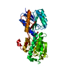
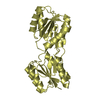
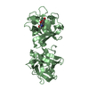
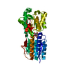
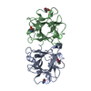
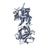
| Title | The crystal structure of a sugar-binding transport protein from Yersinia pestis CO92 | ||||||
 Components Components | Sugar-binding transport protein | ||||||
 Keywords Keywords | SUGAR BINDING PROTEIN /  structural genomics / The Center for Structural Genomics of Infectious Diseases / CSGID / NIAID / National Institute of Allergy and Infectious Diseases structural genomics / The Center for Structural Genomics of Infectious Diseases / CSGID / NIAID / National Institute of Allergy and Infectious Diseases | ||||||
| Function / homology |  Function and homology information Function and homology information | ||||||
| Biological species |   Yersinia pestis (bacteria) Yersinia pestis (bacteria) | ||||||
| Method | X-RAY DIFFRACTION / SYNCHROTRON / SAD / Resolution: 1.65 Å | ||||||
 Authors Authors | Tan, K. / Zhou, M. / Clancy, S. / Anderson, W.F. / Joachimiak, A. / Center for Structural Genomics of Infectious Diseases (CSGID) | ||||||
 Citation Citation | Journal: To be Published Title: The crystal structure of a sugar-binding transport protein from Yersinia pestis CO92 Authors: Tan, K. / Zhou, M. / Clancy, S. / Anderson, W.F. / Joachimiak, A. | ||||||
| History |
|
- Structure visualization
| Structure viewer | Molecule: MolmilJmol/JSmol |
|---|
- Downloads & links
-Download
| PDBx/mmCIF format | 4rwe.cif.gz | 126.9 KB | Display | PDBx/mmCIF format |
|---|---|---|---|---|
| PDB format | pdb4rwe.ent.gz | 103.5 KB | Display | PDB format |
| PDBx/mmJSON format | 4rwe.json.gz | Tree view | PDBx/mmJSON format | |
| Others |  Other downloads Other downloads |
-Validation report
| Arichive directory | https://data.pdbj.org/pub/pdb/validation_reports/rw/4rweftp://data.pdbj.org/pub/pdb/validation_reports/rw/4rwe | HTTPS FTP |
|---|
-Related structure data
| Similar structure data | |
|---|---|
| Other databases |


-Links
 PDBj
PDBj- Assembly
| Deposited unit | 
| ||||||||||||
|---|---|---|---|---|---|---|---|---|---|---|---|---|---|
| 1 |
| ||||||||||||
| Unit cell |
| ||||||||||||
| Components on special symmetry positions |
|
-Components
| #1: Protein | Mass: 30846.275 Da / Num. of mol.: 1 Source method: isolated from a genetically manipulated source Source: (gene. exp.) Yersinia pestis (bacteria) / Strain: CO92 / Gene: A1122_06015, Yersinia pestis / Plasmid: pMCSG68 / Production host: Escherichia coli (E. coli) / Strain (production host): BL21-Gold(DE3) / References: UniProt: G0J922, UniProt: A0A2U2GYC0*PLUS | ||
|---|---|---|---|
| #2: Chemical | ChemComp-GOL / Glycerol  Mass: 92.094 Da / Num. of mol.: 1 / Source method: obtained synthetically / Formula: C3H8O3 Mass: 92.094 Da / Num. of mol.: 1 / Source method: obtained synthetically / Formula: C3H8O3 | ||
| #3: Chemical | Chloride  Mass: 35.453 Da / Num. of mol.: 2 / Source method: obtained synthetically / Formula: Cl Mass: 35.453 Da / Num. of mol.: 2 / Source method: obtained synthetically / Formula: Cl#4: Water | ChemComp-HOH / | Water Mass: 18.015 Da / Num. of mol.: 279 / Source method: isolated from a natural source / Formula: H2O Mass: 18.015 Da / Num. of mol.: 279 / Source method: isolated from a natural source / Formula: H2O |
-Experimental details
-Experiment
| Experiment | Method: X-RAY DIFFRACTION / Number of used crystals: 1 |
|---|
- Sample preparation
| Crystal | Density Matthews: 2.47 Å3/Da / Density % sol: 50.28 % |
|---|---|
| Crystal grow | Temperature: 289 K / Method: vapor diffusion, sitting drop / pH: 7 Details: 1.8M Ammonium citrate tribasic, pH 7.0, VAPOR DIFFUSION, SITTING DROP, temperature 289K |
-Data collection
| Diffraction | Mean temperature: 100 K |
|---|---|
| Diffraction source | Source: SYNCHROTRON / Site: APS  / Beamline: 19-ID / Wavelength: 0.97926 Å / Beamline: 19-ID / Wavelength: 0.97926 Å |
| Detector | Type: ADSC QUANTUM 315r / Detector: CCD / Date: Oct 6, 2014 / Details: Mirror |
| Radiation | Monochromator: Si 111 crystal / Protocol: SINGLE WAVELENGTH / Monochromatic (M) / Laue (L): M / Scattering type: x-ray |
| Radiation wavelength | Wavelength: 0.97926 Å / Relative weight: 1 |
| Reflection | Resolution: 1.65→34.66 Å / Num. all: 37067 / Num. obs: 37067 / % possible obs: 96.8 % / Observed criterion σ(F): 0 / Observed criterion σ(I): -5 / Redundancy: 4.1 % / Biso Wilson estimate: 21.6 Å2 / Rmerge(I) obs: 0.077 / Net I/σ(I): 24.1 |
| Reflection shell | Resolution: 1.65→1.68 Å / Redundancy: 4.2 % / Rmerge(I) obs: 0.764 / Mean I/σ(I) obs: 1.2 / Num. unique all: 1657 / % possible all: 89.2 |
- Processing
| Software |
| ||||||||||||||||||||||||||||||||||||||||||||||||||||||||||||||||||||||||||||||||||||||||||||||||||
|---|---|---|---|---|---|---|---|---|---|---|---|---|---|---|---|---|---|---|---|---|---|---|---|---|---|---|---|---|---|---|---|---|---|---|---|---|---|---|---|---|---|---|---|---|---|---|---|---|---|---|---|---|---|---|---|---|---|---|---|---|---|---|---|---|---|---|---|---|---|---|---|---|---|---|---|---|---|---|---|---|---|---|---|---|---|---|---|---|---|---|---|---|---|---|---|---|---|---|---|
| Refinement | Method to determine structure: SAD / Resolution: 1.65→34.66 Å / SU ML: 0.18 / σ(F): 1.33 / Phase error: 26.17 / Stereochemistry target values: ML
| ||||||||||||||||||||||||||||||||||||||||||||||||||||||||||||||||||||||||||||||||||||||||||||||||||
| Solvent computation | Shrinkage radii: 0.9 Å / VDW probe radii: 1.11 Å / Solvent model: FLAT BULK SOLVENT MODEL | ||||||||||||||||||||||||||||||||||||||||||||||||||||||||||||||||||||||||||||||||||||||||||||||||||
| Refinement step | Cycle: LAST / Resolution: 1.65→34.66 Å
| ||||||||||||||||||||||||||||||||||||||||||||||||||||||||||||||||||||||||||||||||||||||||||||||||||
| Refine LS restraints |
| ||||||||||||||||||||||||||||||||||||||||||||||||||||||||||||||||||||||||||||||||||||||||||||||||||
| LS refinement shell |
|
