 Movie
Movie Controller

[English] 日本語
 Yorodumi
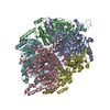
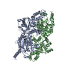
- PDB-4q0g: Crystal structure of beta subunit of acyl-CoA carboxylase AccD1 f... -
+ Open data

- Basic information
| Entry | Database: PDB / ID: 4q0g | ||||||
|---|---|---|---|---|---|---|---|
| Title | Crystal structure of beta subunit of acyl-CoA carboxylase AccD1 from Mycobacterium tuberculosis | ||||||
 Components Components | Probable acetyl-/propionyl-CoA carboxylase (Beta subunit) AccD1 | ||||||
 Keywords Keywords | LIGASE / AccD1 / Structural Genomics / TB Structural Genomics Consortium / TBSGC / crotonase folding / acyl-CoA carboxylase beta subunit / AccA1 (Rv2501c) | ||||||
| Function / homology |  Function and homology information Function and homology informationacyl-CoA biosynthetic process / propionyl-CoA carboxylase / methylcrotonoyl-CoA carboxylase complex / propionyl-CoA carboxylase activity / carboxyl- or carbamoyltransferase activity / L-leucine catabolic process / ligase activity / plasma membrane Similarity search - Function | ||||||
| Biological species |   Mycobacterium tuberculosis (bacteria) Mycobacterium tuberculosis (bacteria) | ||||||
| Method |  X-RAY DIFFRACTION / SYNCHROTRON / MOLECULAR REPLACEMENT / Resolution: 2.31 Å X-RAY DIFFRACTION / SYNCHROTRON / MOLECULAR REPLACEMENT / Resolution: 2.31 Å | ||||||
 Authors Authors | Bie, H.Y. / Yin, J. / James, M.N.G. / TB Structural Genomics Consortium (TBSGC) | ||||||
 Citation Citation | Journal: To be Published Title: Crystal structure of Beta subunit of acyl-CoA carboxylase AccD1 from Mycobacterium tuberculosis Authors: Bie, H.Y. / Yin, J. / James, M.N.G. / TB Structural Genomics Consortium (TBSGC) | ||||||
| History |
|
- Structure visualization
| Structure viewer | Molecule: MolmilJmol/JSmol |
|---|
- Downloads & links
-Download
| PDBx/mmCIF format | 4q0g.cif.gz | 301.9 KB | Display | PDBx/mmCIF format |
|---|---|---|---|---|
| PDB format | pdb4q0g.ent.gz | 244.4 KB | Display | PDB format |
| PDBx/mmJSON format | 4q0g.json.gz | Tree view | PDBx/mmJSON format | |
| Others |  Other downloads Other downloads |
-Validation report
| Summary document | 4q0g_validation.pdf.gz | 448.5 KB | Display | wwPDB validaton report |
|---|---|---|---|---|
| Full document | 4q0g_full_validation.pdf.gz | 455.4 KB | Display | |
| Data in XML | 4q0g_validation.xml.gz | 55.8 KB | Display | |
| Data in CIF | 4q0g_validation.cif.gz | 79.5 KB | Display | |
| Arichive directory | https://data.pdbj.org/pub/pdb/validation_reports/q0/4q0gftp://data.pdbj.org/pub/pdb/validation_reports/q0/4q0g | HTTPS FTP |
-Related structure data
| Related structure data | 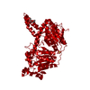 3u9rS S: Starting model for refinement |
|---|---|
| Similar structure data |


-Links
 PDBj
PDBj- Assembly
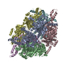
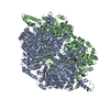
| Deposited unit | 
| ||||||||
|---|---|---|---|---|---|---|---|---|---|
| 1 | 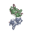
| ||||||||
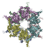
| Unit cell |
|
-Components
| #1: Protein | Mass: 56843.574 Da / Num. of mol.: 3 Source method: isolated from a genetically manipulated source Source: (gene. exp.) Mycobacterium tuberculosis (bacteria) / Gene: pccB-3, accD1, MT2577, Rv2502c / Production host: References: UniProt: O06165, UniProt: I6YDK7*PLUS, propionyl-CoA carboxylase #2: Chemical | ChemComp-GOL /   Mass: 92.094 Da / Num. of mol.: 4 / Source method: obtained synthetically / Formula: C3H8O3 Mass: 92.094 Da / Num. of mol.: 4 / Source method: obtained synthetically / Formula: C3H8O3#3: Water | ChemComp-HOH / |  Mass: 18.015 Da / Num. of mol.: 573 / Source method: isolated from a natural source / Formula: H2O Mass: 18.015 Da / Num. of mol.: 573 / Source method: isolated from a natural source / Formula: H2O |
|---|
-Experimental details
-Experiment
| Experiment | Method: X-RAY DIFFRACTION / Number of used crystals: 1 |
|---|
- Sample preparation
| Crystal | Density Matthews: 3.88 Å3/Da / Density % sol: 68.32 % |
|---|---|
| Crystal grow | Temperature: 298 K / Method: vapor diffusion, hanging drop / pH: 7.5 Details: 0.1 M sodium HEPES pH 7.5 and 30% PEG300, VAPOR DIFFUSION, HANGING DROP, temperature 298K |
-Data collection
| Diffraction | Mean temperature: 110 K |
|---|---|
| Diffraction source | Source: SYNCHROTRON / Site: CLSI  / Beamline: 08ID-1 / Wavelength: 0.97949 Å / Beamline: 08ID-1 / Wavelength: 0.97949 Å |
| Detector | Type: RAYONIX MX300HE / Detector: CCD / Date: Jun 5, 2011 Details: vertical focusing mirror: ultra-low expansion titanium silicate flat mirror with Pt, uncoated, and Pd strips. |
| Radiation | Monochromator: double crystal Si (111) / Protocol: SINGLE WAVELENGTH / Monochromatic (M) / Laue (L): M / Scattering type: x-ray |
| Radiation wavelength | Wavelength: 0.97949 Å / Relative weight: 1 |
| Reflection | Resolution: 2.3→48.81 Å / Num. all: 116259 / Num. obs: 115910 / % possible obs: 99.7 % / Observed criterion σ(F): 0 / Observed criterion σ(I): -3 / Redundancy: 5.8 % / Rmerge(I) obs: 0.157 / Net I/σ(I): 12.93 |
| Reflection shell | Resolution: 2.3→2.38 Å / Redundancy: 5.7 % / Rmerge(I) obs: 0.817 / Mean I/σ(I) obs: 2.66 / Num. unique all: 11512 / % possible all: 100 |
- Processing
| Software |
| |||||||||||||||||||||||||||||||||||||||||||||||||||||||||||||||||||||||||||||||||||||||||||||||||||||||||
|---|---|---|---|---|---|---|---|---|---|---|---|---|---|---|---|---|---|---|---|---|---|---|---|---|---|---|---|---|---|---|---|---|---|---|---|---|---|---|---|---|---|---|---|---|---|---|---|---|---|---|---|---|---|---|---|---|---|---|---|---|---|---|---|---|---|---|---|---|---|---|---|---|---|---|---|---|---|---|---|---|---|---|---|---|---|---|---|---|---|---|---|---|---|---|---|---|---|---|---|---|---|---|---|---|---|---|
| Refinement | Method to determine structure: MOLECULAR REPLACEMENT Starting model: 3U9R Resolution: 2.31→48.81 Å / Cor.coef. Fo:Fc: 0.941 / Cor.coef. Fo:Fc free: 0.93 / SU B: 4.405 / SU ML: 0.106 / Cross valid method: THROUGHOUT / σ(I): -3 / ESU R: 0.2 / ESU R Free: 0.168 / Stereochemistry target values: MAXIMUM LIKELIHOOD / Details: HYDROGENS HAVE BEEN ADDED IN THE RIDING POSITIONS
| |||||||||||||||||||||||||||||||||||||||||||||||||||||||||||||||||||||||||||||||||||||||||||||||||||||||||
| Solvent computation | Ion probe radii: 0.8 Å / Shrinkage radii: 0.8 Å / VDW probe radii: 1.2 Å / Solvent model: MASK | |||||||||||||||||||||||||||||||||||||||||||||||||||||||||||||||||||||||||||||||||||||||||||||||||||||||||
| Displacement parameters | Biso mean: 27.757 Å2
| |||||||||||||||||||||||||||||||||||||||||||||||||||||||||||||||||||||||||||||||||||||||||||||||||||||||||
| Refinement step | Cycle: LAST / Resolution: 2.31→48.81 Å
| |||||||||||||||||||||||||||||||||||||||||||||||||||||||||||||||||||||||||||||||||||||||||||||||||||||||||
| Refine LS restraints |
| |||||||||||||||||||||||||||||||||||||||||||||||||||||||||||||||||||||||||||||||||||||||||||||||||||||||||
| LS refinement shell | Resolution: 2.305→2.365 Å / Total num. of bins used: 20
|
