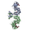
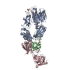
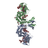
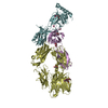
 Movie
Movie Controller


+ Open data

- Basic information
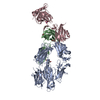
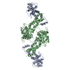
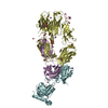
| Entry | Database: PDB / ID: 4pw2 | ||||||
|---|---|---|---|---|---|---|---|
| Title | Crystal structure of D-glucuronyl C5 epimerase | ||||||
 Components Components | D-glucuronyl C5 epimerase B | ||||||
 Keywords Keywords |  ISOMERASE / epimerization enzyme / multiple domain structure / Heparan sulfate C5-epimerase / heparin / heparan sulfate ISOMERASE / epimerization enzyme / multiple domain structure / Heparan sulfate C5-epimerase / heparin / heparan sulfate | ||||||
| Function / homology |  Function and homology informationheparosan-N-sulfate-glucuronate 5-epimerase / heparosan-N-sulfate-glucuronate 5-epimerase activity / heparin biosynthetic process / heparan sulfate proteoglycan biosynthetic process / Golgi membrane / Golgi apparatus / identical protein binding / metal ion binding Function and homology informationheparosan-N-sulfate-glucuronate 5-epimerase / heparosan-N-sulfate-glucuronate 5-epimerase activity / heparin biosynthetic process / heparan sulfate proteoglycan biosynthetic process / Golgi membrane / Golgi apparatus / identical protein binding / metal ion bindingSimilarity search - Function | ||||||
| Biological species |  Danio rerio (zebrafish) Danio rerio (zebrafish) | ||||||
| Method | X-RAY DIFFRACTION / SYNCHROTRON / SAD / Resolution: 1.9 Å | ||||||
 Authors Authors | Ke, J. / Qin, Y. / Gu, X. / Brunzelle, J.S. / Xu, H.E. / Ding, K. | ||||||
 Citation Citation | Journal: J.Biol.Chem. / Year: 2015 Title: Structural and Functional Study of d-Glucuronyl C5-epimerase. Authors: Qin, Y. / Ke, J. / Gu, X. / Fang, J. / Wang, W. / Cong, Q. / Li, J. / Tan, J. / Brunzelle, J.S. / Zhang, C. / Jiang, Y. / Melcher, K. / Li, J.P. / Xu, H.E. / Ding, K. | ||||||
| History |
|
- Structure visualization
| Structure viewer | Molecule: MolmilJmol/JSmol |
|---|
- Downloads & links
-Download
| PDBx/mmCIF format | 4pw2.cif.gz | 130.4 KB | Display | PDBx/mmCIF format |
|---|---|---|---|---|
| PDB format | pdb4pw2.ent.gz | 100.1 KB | Display | PDB format |
| PDBx/mmJSON format | 4pw2.json.gz | Tree view | PDBx/mmJSON format | |
| Others |  Other downloads Other downloads |
-Validation report
| Arichive directory | https://data.pdbj.org/pub/pdb/validation_reports/pw/4pw2ftp://data.pdbj.org/pub/pdb/validation_reports/pw/4pw2 | HTTPS FTP |
|---|
-Related structure data


-Links
 PDBj
PDBj- Assembly
| Deposited unit | 
| |||||||||
|---|---|---|---|---|---|---|---|---|---|---|
| 1 | 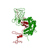
| |||||||||
| Unit cell |
| |||||||||
| Components on special symmetry positions |
|
-Components
| #1: Protein | Mass: 66136.188 Da / Num. of mol.: 1 Source method: isolated from a genetically manipulated source Source: (gene. exp.) Danio rerio (zebrafish) / Gene: D-glucuronyl C5-epimerase, glce, glceb / Plasmid: pSUMO / Production host:  Escherichia coli (E. coli) / Strain (production host): BL21(DE3) Escherichia coli (E. coli) / Strain (production host): BL21(DE3)References: UniProt: Q6TS32, UniProt: F1QR43*PLUS, heparosan-N-sulfate-glucuronate 5-epimerase |
|---|---|
| #2: Chemical | ChemComp-CIT / Citric acid  Mass: 192.124 Da / Num. of mol.: 1 / Source method: obtained synthetically / Formula: C6H8O7 Mass: 192.124 Da / Num. of mol.: 1 / Source method: obtained synthetically / Formula: C6H8O7 |
| #3: Water | ChemComp-HOH / Water Mass: 18.015 Da / Num. of mol.: 684 / Source method: isolated from a natural source / Formula: H2O Mass: 18.015 Da / Num. of mol.: 684 / Source method: isolated from a natural source / Formula: H2O |
-Experimental details
-Experiment
| Experiment | Method: X-RAY DIFFRACTION / Number of used crystals: 1 |
|---|
- Sample preparation
| Crystal | Density Matthews: 2.81 Å3/Da / Density % sol: 56.25 % |
|---|---|
| Crystal grow | Temperature: 293 K / Method: vapor diffusion, sitting drop / pH: 5.6 Details: 16% w/v PEG 3,350, 0.1 M sodium citrate tribasic dihydrate pH 5.6, and 2% v/v Tacsimate pH 5.0, VAPOR DIFFUSION, SITTING DROP, temperature 293K |
-Data collection
| Diffraction | Mean temperature: 100 K |
|---|---|
| Diffraction source | Source: SYNCHROTRON / Site: APS  / Beamline: 21-ID-G / Wavelength: 0.97856 Å / Beamline: 21-ID-G / Wavelength: 0.97856 Å |
| Detector | Type: MARMOSAIC 300 mm CCD / Detector: CCD / Date: Feb 15, 2013 |
| Radiation | Monochromator: Ni FILTER / Protocol: SINGLE WAVELENGTH / Monochromatic (M) / Laue (L): M / Scattering type: x-ray |
| Radiation wavelength | Wavelength: 0.97856 Å / Relative weight: 1 |
| Reflection | Resolution: 1.9→50 Å / Num. all: 61140 / Num. obs: 61140 / % possible obs: 100 % / Redundancy: 14 % / Rmerge(I) obs: 0.101 / Net I/σ(I): 14.8 |
| Reflection shell | Resolution: 1.9→2 Å / Redundancy: 14.7 % / Rmerge(I) obs: 1.04 / Mean I/σ(I) obs: 3.2 / Num. unique all: 8707 / % possible all: 99.9 |
- Processing
| Software |
| |||||||||||||||||||||||||||||||||||||||||||||||||||||||||||||||||
|---|---|---|---|---|---|---|---|---|---|---|---|---|---|---|---|---|---|---|---|---|---|---|---|---|---|---|---|---|---|---|---|---|---|---|---|---|---|---|---|---|---|---|---|---|---|---|---|---|---|---|---|---|---|---|---|---|---|---|---|---|---|---|---|---|---|---|
| Refinement | Method to determine structure: SAD / Resolution: 1.9→47.38 Å / Cor.coef. Fo:Fc: 0.955 / Cor.coef. Fo:Fc free: 0.936 / SU B: 3.829 / SU ML: 0.108 / Cross valid method: THROUGHOUT / ESU R: 0.143 / ESU R Free: 0.136 / Stereochemistry target values: MAXIMUM LIKELIHOOD
| |||||||||||||||||||||||||||||||||||||||||||||||||||||||||||||||||
| Solvent computation | Ion probe radii: 0.8 Å / Shrinkage radii: 0.8 Å / VDW probe radii: 1.2 Å / Solvent model: MASK | |||||||||||||||||||||||||||||||||||||||||||||||||||||||||||||||||
| Displacement parameters | Biso mean: 36.239 Å2
| |||||||||||||||||||||||||||||||||||||||||||||||||||||||||||||||||
| Refinement step | Cycle: LAST / Resolution: 1.9→47.38 Å
| |||||||||||||||||||||||||||||||||||||||||||||||||||||||||||||||||
| Refine LS restraints |
| |||||||||||||||||||||||||||||||||||||||||||||||||||||||||||||||||
| LS refinement shell | Resolution: 1.9→1.949 Å / Total num. of bins used: 20
|
