 Movie
Movie Controller


+ Open data

- Basic information
| Entry | Database: PDB / ID: 4n4r | ||||||
|---|---|---|---|---|---|---|---|
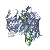
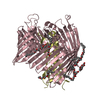
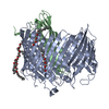
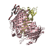
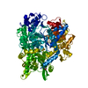
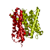
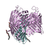
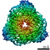
| Title | Structure basis of lipopolysaccharide biogenesis | ||||||
 Components Components |
| ||||||
 Keywords Keywords |  MEMBRANE PROTEIN / Beta barrel / Translocase / Lipopolysaccharide transport proteins MEMBRANE PROTEIN / Beta barrel / Translocase / Lipopolysaccharide transport proteins | ||||||
| Function / homology |  Function and homology information Function and homology informationlipopolysaccharide transport => GO:0015920 / lipopolysaccharide transport / Gram-negative-bacterium-type cell outer membrane assembly / response to organic substance / lipopolysaccharide binding / cell outer membraneSimilarity search - Function | ||||||
| Biological species |  Salmonella enterica subsp. enterica serovar Typhimurium (bacteria) Salmonella enterica subsp. enterica serovar Typhimurium (bacteria) | ||||||
| Method | X-RAY DIFFRACTION / SYNCHROTRON / MAD / Resolution: 2.8 Å | ||||||
 Authors Authors | Dong, H. / Xiang, Q. / Wang, Z. / Paterson, N.G. / He, C. / Zhang, Y. / Wang, W. / Dong, C. | ||||||
 Citation Citation | Journal: Nature / Year: 2014 Title: Structural basis for outer membrane lipopolysaccharide insertion. Authors: Dong, H. / Xiang, Q. / Gu, Y. / Wang, Z. / Paterson, N.G. / Stansfeld, P.J. / He, C. / Zhang, Y. / Wang, W. / Dong, C. | ||||||
| History |
|
- Structure visualization
| Structure viewer | Molecule: MolmilJmol/JSmol |
|---|
- Downloads & links
-Download
| PDBx/mmCIF format | 4n4r.cif.gz | 575.1 KB | Display | PDBx/mmCIF format |
|---|---|---|---|---|
| PDB format | pdb4n4r.ent.gz | 494.1 KB | Display | PDB format |
| PDBx/mmJSON format | 4n4r.json.gz | Tree view | PDBx/mmJSON format | |
| Others |  Other downloads Other downloads |
-Validation report
| Arichive directory | https://data.pdbj.org/pub/pdb/validation_reports/n4/4n4rftp://data.pdbj.org/pub/pdb/validation_reports/n4/4n4r | HTTPS FTP |
|---|
-Related structure data
| Similar structure data |
|---|

-Links
 PDBj
PDBj


- Assembly
| Deposited unit | 
| ||||||||||||||||||||||||||||||||||||||||||||||||
|---|---|---|---|---|---|---|---|---|---|---|---|---|---|---|---|---|---|---|---|---|---|---|---|---|---|---|---|---|---|---|---|---|---|---|---|---|---|---|---|---|---|---|---|---|---|---|---|---|---|
| 1 | 
| ||||||||||||||||||||||||||||||||||||||||||||||||
| 2 | 
| ||||||||||||||||||||||||||||||||||||||||||||||||
| Unit cell |
| ||||||||||||||||||||||||||||||||||||||||||||||||
| Noncrystallographic symmetry (NCS) | NCS domain:
NCS domain segments:
NCS ensembles :
|
-Components
| #1: Protein | Mass: 90852.211 Da / Num. of mol.: 2 Source method: isolated from a genetically manipulated source Source: (gene. exp.) Salmonella enterica subsp. enterica serovar Typhimurium (bacteria)Strain: LT2/ SGSC1412 / ATCC 700720 / Gene: imp, lptD, ostA, STM0093 / Plasmid: pET28a / Production host: Escherichia coli (E. coli) / Strain (production host): C43 / References: UniProt: Q8ZRW0#2: Protein | Mass: 21853.621 Da / Num. of mol.: 2 Source method: isolated from a genetically manipulated source Source: (gene. exp.) Salmonella enterica subsp. enterica serovar Typhimurium (bacteria)Strain: LT2 / SGSC1412 / ATCC 700720 / Gene: lptE, rlpB, STM0647 / Plasmid: pACYC-Duet-1 / Production host: Escherichia coli (E. coli) / Strain (production host): C43 / References: UniProt: Q8ZQZ7#3: Chemical | Cacodylic acid  Mass: 136.989 Da / Num. of mol.: 2 / Source method: obtained synthetically / Formula: C2H6AsO2 Mass: 136.989 Da / Num. of mol.: 2 / Source method: obtained synthetically / Formula: C2H6AsO2#4: Chemical |   Mass: 65.409 Da / Num. of mol.: 2 / Source method: obtained synthetically / Formula: Zn Mass: 65.409 Da / Num. of mol.: 2 / Source method: obtained synthetically / Formula: Zn#5: Water | ChemComp-HOH / | Water Mass: 18.015 Da / Num. of mol.: 10 / Source method: isolated from a natural source / Formula: H2O Mass: 18.015 Da / Num. of mol.: 10 / Source method: isolated from a natural source / Formula: H2O |
|---|
-Experimental details
-Experiment
| Experiment | Method: X-RAY DIFFRACTION / Number of used crystals: 3 |
|---|
- Sample preparation
| Crystal | Density Matthews: 2.91 Å3/Da / Density % sol: 57.7 % |
|---|---|
| Crystal grow | Temperature: 293 K / Method: vapor diffusion, sitting drop / pH: 7 Details: 0.1 M zinc acetate, 0.1M sodium cacodylate, 18% PEG8000 , pH 7.0, VAPOR DIFFUSION, SITTING DROP, temperature 293K |
-Data collection
| Diffraction | Mean temperature: 273 K | |||||||||||||||
|---|---|---|---|---|---|---|---|---|---|---|---|---|---|---|---|---|
| Diffraction source | Source: SYNCHROTRON / Site: Diamond  / Beamline: I24 / Wavelength: 0.97887, 0.97835, 0.98181, 0.97750 / Beamline: I24 / Wavelength: 0.97887, 0.97835, 0.98181, 0.97750 | |||||||||||||||
| Detector | Type: DECTRIS PILATUS 6M / Detector: PIXEL / Date: May 24, 2013 | |||||||||||||||
| Radiation | Monochromator: Graphite / Protocol: MAD / Monochromatic (M) / Laue (L): M / Scattering type: x-ray | |||||||||||||||
| Radiation wavelength |
| |||||||||||||||
| Reflection | Resolution: 2.8→107.49 Å / Num. obs: 64252 / % possible obs: 9.7 % / Observed criterion σ(F): 2 / Observed criterion σ(I): 1.6 / Redundancy: 5.6 % / Rmerge(I) obs: 0.154 / Rsym value: 0.16 / Net I/σ(I): 1.6 | |||||||||||||||
| Reflection shell | Resolution: 2.8→3 Å / Redundancy: 4.8 % / Rmerge(I) obs: 0.904 / Mean I/σ(I) obs: 1.6 / Rsym value: 0.93 / % possible all: 94 |
- Processing
| Software |
| ||||||||||||||||||||||||||||||||||||||||||||||||||||||||||||||||||||||||||||||||||||||||||||||||||||||||||||||||||||||||||||||||||||||||||||||||||||||||||||||||||||||||||||||||||||||
|---|---|---|---|---|---|---|---|---|---|---|---|---|---|---|---|---|---|---|---|---|---|---|---|---|---|---|---|---|---|---|---|---|---|---|---|---|---|---|---|---|---|---|---|---|---|---|---|---|---|---|---|---|---|---|---|---|---|---|---|---|---|---|---|---|---|---|---|---|---|---|---|---|---|---|---|---|---|---|---|---|---|---|---|---|---|---|---|---|---|---|---|---|---|---|---|---|---|---|---|---|---|---|---|---|---|---|---|---|---|---|---|---|---|---|---|---|---|---|---|---|---|---|---|---|---|---|---|---|---|---|---|---|---|---|---|---|---|---|---|---|---|---|---|---|---|---|---|---|---|---|---|---|---|---|---|---|---|---|---|---|---|---|---|---|---|---|---|---|---|---|---|---|---|---|---|---|---|---|---|---|---|---|---|
| Refinement | Method to determine structure: MAD / Resolution: 2.8→107.49 Å / Cor.coef. Fo:Fc: 0.892 / Cor.coef. Fo:Fc free: 0.907 / SU B: 44.165 / SU ML: 0.371 / Cross valid method: THROUGHOUT / ESU R: 0.667 / ESU R Free: 0.371 / Stereochemistry target values: MAXIMUM LIKELIHOOD / Details: HYDROGENS HAVE BEEN USED IF PRESENT IN THE INPUT
| ||||||||||||||||||||||||||||||||||||||||||||||||||||||||||||||||||||||||||||||||||||||||||||||||||||||||||||||||||||||||||||||||||||||||||||||||||||||||||||||||||||||||||||||||||||||
| Solvent computation | Ion probe radii: 0.8 Å / Shrinkage radii: 0.8 Å / VDW probe radii: 1.2 Å / Solvent model: MASK | ||||||||||||||||||||||||||||||||||||||||||||||||||||||||||||||||||||||||||||||||||||||||||||||||||||||||||||||||||||||||||||||||||||||||||||||||||||||||||||||||||||||||||||||||||||||
| Displacement parameters | Biso mean: 120.272 Å2
| ||||||||||||||||||||||||||||||||||||||||||||||||||||||||||||||||||||||||||||||||||||||||||||||||||||||||||||||||||||||||||||||||||||||||||||||||||||||||||||||||||||||||||||||||||||||
| Refine analyze |
| ||||||||||||||||||||||||||||||||||||||||||||||||||||||||||||||||||||||||||||||||||||||||||||||||||||||||||||||||||||||||||||||||||||||||||||||||||||||||||||||||||||||||||||||||||||||
| Refinement step | Cycle: LAST / Resolution: 2.8→107.49 Å
| ||||||||||||||||||||||||||||||||||||||||||||||||||||||||||||||||||||||||||||||||||||||||||||||||||||||||||||||||||||||||||||||||||||||||||||||||||||||||||||||||||||||||||||||||||||||
| Refine LS restraints |
|