 Movie
Movie Controller

[English] 日本語
 Yorodumi
- PDB-4avo: Thermobifida fusca cellobiohydrolase Cel6B catalytic mutant D274A... -
+ Open data

- Basic information
| Entry | Database: PDB / ID: 4avo | |||||||||
|---|---|---|---|---|---|---|---|---|---|---|
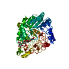
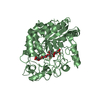
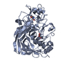
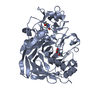
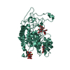
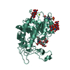
| Title | Thermobifida fusca cellobiohydrolase Cel6B catalytic mutant D274A cocrystallized with cellobiose | |||||||||
 Components Components | BETA-1,4-EXOCELLULASE | |||||||||
 Keywords Keywords | HYDROLASE / CELLULOSE DEGRADATION / GLYCOSIDE HYDROLASE FAMILY 6 / CELLULASE | |||||||||
| Function / homology |  Function and homology information Function and homology informationHydrolases; Glycosylases; Glycosidases, i.e. enzymes that hydrolyse O- and S-glycosyl compounds / polysaccharide binding / cellulose catabolic process / hydrolase activity, hydrolyzing O-glycosyl compounds / metal ion binding Similarity search - Function | |||||||||
| Biological species |   THERMOBIFIDA FUSCA (bacteria) THERMOBIFIDA FUSCA (bacteria) | |||||||||
| Method |  X-RAY DIFFRACTION / SYNCHROTRON / MOLECULAR REPLACEMENT / Resolution: 1.8 Å X-RAY DIFFRACTION / SYNCHROTRON / MOLECULAR REPLACEMENT / Resolution: 1.8 Å | |||||||||
 Authors Authors | Wu, M. / Vuong, T.V. / Wilson, D.B. / Sandgren, M. / Stahlberg, J. / Hansson, H. | |||||||||
 Citation Citation | Journal: J.Biol.Chem. / Year: 2013 Title: Loop Motions Important to Product Expulsion in the Thermobifida Fusca Glycoside Hydrolase Family 6 Cellobiohydrolase from Structural and Computational Studies. Authors: Wu, M. / Bu, L. / Vuong, T.V. / Wilson, D.B. / Crowley, M.F. / Sandgren, M. / Stahlberg, J. / Beckham, G.T. / Hansson, H. | |||||||||
| History |
|
- Structure visualization
| Structure viewer | Molecule: MolmilJmol/JSmol |
|---|
- Downloads & links
-Download
| PDBx/mmCIF format | 4avo.cif.gz | 104.1 KB | Display | PDBx/mmCIF format |
|---|---|---|---|---|
| PDB format | pdb4avo.ent.gz | 77.5 KB | Display | PDB format |
| PDBx/mmJSON format | 4avo.json.gz | Tree view | PDBx/mmJSON format | |
| Others |  Other downloads Other downloads |
-Validation report
| Arichive directory | https://data.pdbj.org/pub/pdb/validation_reports/av/4avoftp://data.pdbj.org/pub/pdb/validation_reports/av/4avo | HTTPS FTP |
|---|
-Related structure data


-Links
 PDBj
PDBj


- Assembly
| Deposited unit | 
| ||||||||
|---|---|---|---|---|---|---|---|---|---|
| 1 |
| ||||||||
| Unit cell |
|
-Components
| #1: Protein | Mass: 45429.738 Da / Num. of mol.: 1 / Fragment: CATALYTIC DOMAIN, RESIDUES 177-596 / Mutation: YES Source method: isolated from a genetically manipulated source Source: (gene. exp.) THERMOBIFIDA FUSCA (bacteria) / Strain: YX / Production host: References: UniProt: Q60029, UniProt: Q47SA9*PLUS, cellulose 1,4-beta-cellobiosidase (non-reducing end) |
|---|---|
| #2: Polysaccharide | beta-D-glucopyranose-(1-4)-beta-D-glucopyranose-(1-4)-beta-D-glucopyranose-(1-4)-beta-D- ...beta-D-glucopyranose-(1-4)-beta-D-glucopyranose-(1-4)-beta-D-glucopyranose-(1-4)-beta-D-glucopyranose-(1-4)-beta-D-glucopyranose-(1-4)-beta-D-glucopyranose / beta-cellohexaose  Source method: isolated from a genetically manipulated source Details: oligosaccharide / References: beta-cellohexaose |
| #3: Chemical | ChemComp-CA /   Mass: 40.078 Da / Num. of mol.: 1 / Source method: obtained synthetically / Formula: Ca Mass: 40.078 Da / Num. of mol.: 1 / Source method: obtained synthetically / Formula: Ca |
| #4: Chemical | ChemComp-ACT /   Mass: 59.044 Da / Num. of mol.: 1 / Source method: obtained synthetically / Formula: C2H3O2 Mass: 59.044 Da / Num. of mol.: 1 / Source method: obtained synthetically / Formula: C2H3O2 |
| #5: Water | ChemComp-HOH /  Mass: 18.015 Da / Num. of mol.: 304 / Source method: isolated from a natural source / Formula: H2O Mass: 18.015 Da / Num. of mol.: 304 / Source method: isolated from a natural source / Formula: H2O |
| Has protein modification | Y |
-Experimental details
-Experiment
| Experiment | Method: X-RAY DIFFRACTION / Number of used crystals: 1 |
|---|
- Sample preparation
| Crystal | Density Matthews: 2.01 Å3/Da / Density % sol: 38.84 % / Description: NONE |
|---|---|
| Crystal grow | pH: 4 Details: 20% PEG 6000, 0.1 M CACL2, 0.1 M SODIUM ACETATE PH 4, ABOUT 10MM CELLOBIOSE. |
-Data collection
| Diffraction | Mean temperature: 100 K |
|---|---|
| Diffraction source | Source: SYNCHROTRON / Site: ESRF  / Beamline: ID23-1 / Wavelength: 0.97685 / Beamline: ID23-1 / Wavelength: 0.97685 |
| Detector | Type: ADSC QUANTUM 315r / Detector: CCD / Date: Sep 17, 2010 |
| Radiation | Monochromator: SI(111) / Protocol: SINGLE WAVELENGTH / Monochromatic (M) / Laue (L): M / Scattering type: x-ray |
| Radiation wavelength | Wavelength: 0.97685 Å / Relative weight: 1 |
| Reflection | Resolution: 1.75→46.1 Å / Num. obs: 32986 / % possible obs: 99.9 % / Observed criterion σ(I): 2.3 / Redundancy: 4.1 % / Rmerge(I) obs: 0.1 / Net I/σ(I): 10.7 |
| Reflection shell | Resolution: 1.8→1.9 Å / Redundancy: 4.1 % / Rmerge(I) obs: 0.3 / Mean I/σ(I) obs: 4.9 / % possible all: 99.9 |
- Processing
| Software |
| ||||||||||||||||||||||||||||||||||||||||||||||||||||||||||||||||||||||||||||||||||||||||||||||||||||||||||||||||||||||||||||||||||||||||||||||||||||||||||||||||||||||||||||||||||||||
|---|---|---|---|---|---|---|---|---|---|---|---|---|---|---|---|---|---|---|---|---|---|---|---|---|---|---|---|---|---|---|---|---|---|---|---|---|---|---|---|---|---|---|---|---|---|---|---|---|---|---|---|---|---|---|---|---|---|---|---|---|---|---|---|---|---|---|---|---|---|---|---|---|---|---|---|---|---|---|---|---|---|---|---|---|---|---|---|---|---|---|---|---|---|---|---|---|---|---|---|---|---|---|---|---|---|---|---|---|---|---|---|---|---|---|---|---|---|---|---|---|---|---|---|---|---|---|---|---|---|---|---|---|---|---|---|---|---|---|---|---|---|---|---|---|---|---|---|---|---|---|---|---|---|---|---|---|---|---|---|---|---|---|---|---|---|---|---|---|---|---|---|---|---|---|---|---|---|---|---|---|---|---|---|
| Refinement | Method to determine structure: MOLECULAR REPLACEMENT Starting model: UNPUBLISHED WILD TYPE CEL6B Resolution: 1.8→20 Å / Cor.coef. Fo:Fc: 0.958 / Cor.coef. Fo:Fc free: 0.938 / SU B: 2.183 / SU ML: 0.07 / Cross valid method: THROUGHOUT / ESU R: 0.133 / ESU R Free: 0.12 / Stereochemistry target values: MAXIMUM LIKELIHOOD Details: HYDROGENS HAVE BEEN ADDED IN THE RIDING POSITIONS. U VALUES REFINED INDIVIDUALLY.
| ||||||||||||||||||||||||||||||||||||||||||||||||||||||||||||||||||||||||||||||||||||||||||||||||||||||||||||||||||||||||||||||||||||||||||||||||||||||||||||||||||||||||||||||||||||||
| Solvent computation | Ion probe radii: 0.8 Å / Shrinkage radii: 0.8 Å / VDW probe radii: 1.4 Å / Solvent model: MASK | ||||||||||||||||||||||||||||||||||||||||||||||||||||||||||||||||||||||||||||||||||||||||||||||||||||||||||||||||||||||||||||||||||||||||||||||||||||||||||||||||||||||||||||||||||||||
| Displacement parameters | Biso mean: 11.995 Å2
| ||||||||||||||||||||||||||||||||||||||||||||||||||||||||||||||||||||||||||||||||||||||||||||||||||||||||||||||||||||||||||||||||||||||||||||||||||||||||||||||||||||||||||||||||||||||
| Refinement step | Cycle: LAST / Resolution: 1.8→20 Å
| ||||||||||||||||||||||||||||||||||||||||||||||||||||||||||||||||||||||||||||||||||||||||||||||||||||||||||||||||||||||||||||||||||||||||||||||||||||||||||||||||||||||||||||||||||||||
| Refine LS restraints |
|