| 登録情報 | データベース: PDB / ID: 3pqr
|
|---|
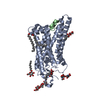
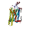
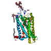
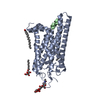
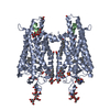
| タイトル | Crystal structure of Metarhodopsin II in complex with a C-terminal peptide derived from the Galpha subunit of transducin |
|---|
 要素 要素 | - Guanine nucleotide-binding protein G(t) subunit alpha-1
- Rhodopsin
|
|---|
 キーワード キーワード | SIGNALING PROTEIN / protein / retinal protein / photoreceptor / active state / chromophore / G-protein coupled receptor / glycoprotein / lipoprotein / palmitate / phosphoprotein / photoreceptor protein / sensory transduction / transducer / transmembrane / vision / G-protein / transducin / Galpha subunit / membrane / receptor / GTP-binding / myristate / nucleotide-binding / G-protein-coupled receptor / rhodopsin / opsin |
|---|
| 機能・相同性 |  機能・相同性情報 機能・相同性情報
negative regulation of cyclic-nucleotide phosphodiesterase activity / Opsins / VxPx cargo-targeting to cilium / rod bipolar cell differentiation / sperm head plasma membrane / absorption of visible light / opsin binding / The canonical retinoid cycle in rods (twilight vision) / G protein-coupled opsin signaling pathway / 11-cis retinal binding ...negative regulation of cyclic-nucleotide phosphodiesterase activity / Opsins / VxPx cargo-targeting to cilium / rod bipolar cell differentiation / sperm head plasma membrane / absorption of visible light / opsin binding / The canonical retinoid cycle in rods (twilight vision) / G protein-coupled opsin signaling pathway / 11-cis retinal binding / podosome assembly / G protein-coupled photoreceptor activity / photoreceptor inner segment membrane / cellular response to light stimulus / rod photoreceptor outer segment / detection of light stimulus involved in visual perception / G protein-coupled receptor complex / Inactivation, recovery and regulation of the phototransduction cascade / thermotaxis / Activation of the phototransduction cascade / outer membrane / detection of temperature stimulus involved in thermoception / response to light intensity / photoreceptor cell maintenance / arrestin family protein binding / photoreceptor outer segment membrane / G alpha (i) signalling events / acyl binding / response to light stimulus / phototransduction, visible light / phototransduction / G-protein alpha-subunit binding / photoreceptor outer segment / photoreceptor inner segment / visual perception / guanyl-nucleotide exchange factor activity / G protein-coupled receptor binding / microtubule cytoskeleton organization / adenylate cyclase-modulating G protein-coupled receptor signaling pathway / G-protein beta/gamma-subunit complex binding / cell-cell junction / photoreceptor disc membrane / GDP binding / sperm midpiece / heterotrimeric G-protein complex / gene expression / G protein-coupled receptor signaling pathway / Golgi membrane / GTPase activity / protein kinase binding / GTP binding / zinc ion binding / metal ion binding / identical protein binding / membrane / plasma membrane / cytoplasm類似検索 - 分子機能 Rhodopsin, N-terminal / Amino terminal of the G-protein receptor rhodopsin / Rhodopsin / Opsin / Visual pigments (opsins) retinal binding site / Visual pigments (opsins) retinal binding site. / : / Rhopdopsin 7-helix transmembrane proteins / Rhodopsin 7-helix transmembrane proteins / G-protein alpha subunit, group I ...Rhodopsin, N-terminal / Amino terminal of the G-protein receptor rhodopsin / Rhodopsin / Opsin / Visual pigments (opsins) retinal binding site / Visual pigments (opsins) retinal binding site. / : / Rhopdopsin 7-helix transmembrane proteins / Rhodopsin 7-helix transmembrane proteins / G-protein alpha subunit, group I / Serpentine type 7TM GPCR chemoreceptor Srsx / Guanine nucleotide binding protein (G-protein), alpha subunit / G protein alpha subunit, helical insertion / G-protein alpha subunit / G-alpha domain profile. / G protein alpha subunit / G-protein coupled receptors family 1 signature. / 7 transmembrane receptor (rhodopsin family) / G protein-coupled receptor, rhodopsin-like / GPCR, rhodopsin-like, 7TM / G-protein coupled receptors family 1 profile. / Up-down Bundle / P-loop containing nucleoside triphosphate hydrolase / Mainly Alpha類似検索 - ドメイン・相同性 trehalose / ACETATE ION / PALMITIC ACID / RETINAL / Rhodopsin / Guanine nucleotide-binding protein G(t) subunit alpha-1類似検索 - 構成要素 |
|---|
| 生物種 |   Bos taurus (ウシ) Bos taurus (ウシ) |
|---|
| 手法 |  X線回折 / シンクロトロン / 分子置換 / 解像度: 2.85 Å X線回折 / シンクロトロン / 分子置換 / 解像度: 2.85 Å |
|---|
 データ登録者 データ登録者 | Choe, H.-W. / Kim, Y.J. / Park, J.H. / Morizumi, T. / Pai, E.F. / Krauss, N. / Hofmann, K.P. / Scheerer, P. / Ernst, O.P. |
|---|
 引用 引用 | ジャーナル: Nature / 年: 2011
タイトル: Crystal structure of metarhodopsin II.
著者: Choe, H.W. / Kim, Y.J. / Park, J.H. / Morizumi, T. / Pai, E.F. / Krauss, N. / Hofmann, K.P. / Scheerer, P. / Ernst, O.P. |
|---|
| 履歴 | | 登録 | 2010年11月26日 | 登録サイト: RCSB / 処理サイト: RCSB |
|---|
| 改定 1.0 | 2011年3月9日 | Provider: repository / タイプ: Initial release |
|---|
| 改定 1.1 | 2011年7月13日 | Group: Version format compliance |
|---|
| 改定 2.0 | 2020年7月29日 | Group: Atomic model / Data collection ...Atomic model / Data collection / Database references / Derived calculations / Non-polymer description / Structure summary
カテゴリ: atom_site / chem_comp ...atom_site / chem_comp / entity / entity_name_com / pdbx_branch_scheme / pdbx_chem_comp_identifier / pdbx_entity_branch / pdbx_entity_branch_descriptor / pdbx_entity_branch_link / pdbx_entity_branch_list / pdbx_entity_nonpoly / pdbx_molecule_features / pdbx_nonpoly_scheme / pdbx_struct_assembly_gen / struct_asym / struct_conn / struct_ref_seq_dif / struct_site / struct_site_gen
Item: _atom_site.B_iso_or_equiv / _atom_site.Cartn_x ..._atom_site.B_iso_or_equiv / _atom_site.Cartn_x / _atom_site.Cartn_y / _atom_site.Cartn_z / _atom_site.auth_asym_id / _atom_site.auth_atom_id / _atom_site.auth_comp_id / _atom_site.auth_seq_id / _atom_site.label_asym_id / _atom_site.label_atom_id / _atom_site.label_comp_id / _atom_site.label_entity_id / _atom_site.type_symbol / _chem_comp.formula / _chem_comp.formula_weight / _chem_comp.id / _chem_comp.mon_nstd_flag / _chem_comp.name / _chem_comp.pdbx_synonyms / _chem_comp.type / _pdbx_struct_assembly_gen.asym_id_list / _struct_ref_seq_dif.details
解説: Carbohydrate remediation / Provider: repository / タイプ: Remediation |
|---|
| 改定 2.1 | 2023年9月6日 | Group: Data collection / Database references ...Data collection / Database references / Refinement description / Structure summary
カテゴリ: chem_comp / chem_comp_atom ...chem_comp / chem_comp_atom / chem_comp_bond / database_2 / pdbx_initial_refinement_model
Item: _chem_comp.pdbx_synonyms / _database_2.pdbx_DOI / _database_2.pdbx_database_accession |
|---|
| 改定 2.2 | 2024年10月30日 | Group: Structure summary
カテゴリ: pdbx_entry_details / pdbx_modification_feature |
|---|
|
|---|
 ムービー
ムービー コントローラー


 データを開く
 基本情報
基本情報 構造の表示
構造の表示 ダウンロードとリンク
 その他のダウンロード
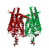


 PDBj
PDBj


 集合体
集合体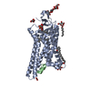


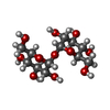

 タイプ: D-saccharide, beta linking / 分子量: 221.208 Da / 分子数: 1 / 由来タイプ: 組換発現 / 式: C8H15NO6
 タイプ: D-saccharide / 分子量: 292.369 Da / 分子数: 2 / 由来タイプ: 組換発現 / 式: C14H28O6 / コメント: 可溶化剤*YM


 分子量: 284.436 Da / 分子数: 1 / 由来タイプ: 合成 / 式: C20H28O
 分子量: 256.424 Da / 分子数: 1 / 由来タイプ: 合成 / 式: C16H32O2
 分子量: 96.063 Da / 分子数: 1 / 由来タイプ: 合成 / 式: SO4
 分子量: 59.044 Da / 分子数: 2 / 由来タイプ: 合成 / 式: C2H3O2
 試料調製
試料調製 / ビームライン: 14.2 / 波長: 0.91841 Å
 解析
解析