- PDB-3oo2: 2.37 Angstrom resolution crystal structure of an alanine racemase... -
+
Open data
ID or keywords:
Loading...
-
Basic information
Entry
Database: PDB / ID: 3oo2
Title
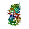
2.37 Angstrom resolution crystal structure of an alanine racemase (alr) from Staphylococcus aureus subsp. aureus COL
Components
Alanine racemase 1
Keywords
ISOMERASE / Alanine Racemase / Infectious Diseases / Center for Structural Genomics of Infectious Diseases (CSGID) / Alanine and Aspartate Metabolism and D-alanine Metabolism / Biosynthesis and degradation of murein sacculus and peptidoglycan
Function / homology
Function and homology information
alanine racemase / D-alanine biosynthetic process / alanine racemase activity / peptidoglycan biosynthetic process / pyridoxal phosphate binding / cytosol Similarity search - Function
Mass: 18.015 Da / Num. of mol.: 120 / Source method: isolated from a natural source / Formula: H2O
-
Experimental details
-
Experiment
Experiment
Method: X-RAY DIFFRACTION / Number of used crystals: 1
-
Sample preparation
Crystal
Density Matthews: 2.11 Å3/Da / Density % sol: 41.73 %
Crystal grow
Temperature: 295 K / Method: vapor diffusion, sitting drop / pH: 5 Details: 7.6 mg/mL protein in 10 mM Tris/HCl pH 8.3, 0.5 M NaCl, 5 mM BME. Crystals grew from 0.1 M MIB buffer pH 5.0, 25 % (w/v) PEG1500 (The PACT suite condition #14), VAPOR DIFFUSION, SITTING DROP, temperature 295K
In the structure databanks used in Yorodumi, some data are registered as the other names, "COVID-19 virus" and "2019-nCoV". Here are the details of the virus and the list of structure data.
Jan 31, 2019. EMDB accession codes are about to change! (news from PDBe EMDB page)
EMDB accession codes are about to change! (news from PDBe EMDB page)
The allocation of 4 digits for EMDB accession codes will soon come to an end. Whilst these codes will remain in use, new EMDB accession codes will include an additional digit and will expand incrementally as the available range of codes is exhausted. The current 4-digit format prefixed with “EMD-” (i.e. EMD-XXXX) will advance to a 5-digit format (i.e. EMD-XXXXX), and so on. It is currently estimated that the 4-digit codes will be depleted around Spring 2019, at which point the 5-digit format will come into force.
The EM Navigator/Yorodumi systems omit the EMD- prefix.
Related info.:Q: What is EMD? / ID/Accession-code notation in Yorodumi/EM Navigator
Yorodumi is a browser for structure data from EMDB, PDB, SASBDB, etc.
This page is also the successor to EM Navigator detail page, and also detail information page/front-end page for Omokage search.
The word "yorodu" (or yorozu) is an old Japanese word meaning "ten thousand". "mi" (miru) is to see.
Related info.:EMDB / PDB / SASBDB / Comparison of 3 databanks / Yorodumi Search / Aug 31, 2016. New EM Navigator & Yorodumi / Yorodumi Papers / Jmol/JSmol / Function and homology information / Changes in new EM Navigator and Yorodumi
 Movie
Movie Controller

 Yorodumi
 Open data
 Basic information
 Components
 Keywords
 Function and homology information
 Staphylococcus aureus subsp. aureus (bacteria)
 X-RAY DIFFRACTION /
  Authors
 Citation
 Structure visualization
 Downloads & links
 Other downloads


 PDBj
PDBj


 Assembly
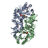

 Mass: 22.990 Da / Num. of mol.: 2 / Source method: obtained synthetically / Formula: Na
 Mass: 78.133 Da / Num. of mol.: 3 / Source method: obtained synthetically / Formula: C2H6OS
 Mass: 94.971 Da / Num. of mol.: 2 / Source method: obtained synthetically / Formula: PO4
 Mass: 18.015 Da / Num. of mol.: 120 / Source method: isolated from a natural source / Formula: H2O
 Sample preparation
 / Beamline: 21-ID-F / Wavelength: 0.97872 Å
 Processing