 Movie
Movie Controller

[English] 日本語
 Yorodumi
- PDB-3kbr: The crystal structure of cyclohexadienyl dehydratase precursor fr... -
+ Open data

- Basic information
| Entry | Database: PDB / ID: 3kbr | ||||||
|---|---|---|---|---|---|---|---|
| Title | The crystal structure of cyclohexadienyl dehydratase precursor from Pseudomonas aeruginosa PA01 | ||||||
 Components Components | Cyclohexadienyl dehydratase | ||||||
 Keywords Keywords |  LYASE / cyclohexadienyl dehydratase precursor / Pseudomonas aeruginosa PA01 / structural genomics / PSI-2 / protein structure initiative / midwest center for structural genomics / MCSG / Amino-acid biosynthesis / Aromatic amino acid biosynthesis / Multifunctional enzyme / Phenylalanine biosynthesis LYASE / cyclohexadienyl dehydratase precursor / Pseudomonas aeruginosa PA01 / structural genomics / PSI-2 / protein structure initiative / midwest center for structural genomics / MCSG / Amino-acid biosynthesis / Aromatic amino acid biosynthesis / Multifunctional enzyme / Phenylalanine biosynthesis | ||||||
| Function / homology |  Function and homology informationarogenate dehydratase / arogenate dehydratase activity / prephenate dehydratase / prephenate dehydratase activity / L-phenylalanine biosynthetic process / periplasmic space Function and homology informationarogenate dehydratase / arogenate dehydratase activity / prephenate dehydratase / prephenate dehydratase activity / L-phenylalanine biosynthetic process / periplasmic spaceSimilarity search - Function | ||||||
| Biological species |   Pseudomonas aeruginosa (bacteria) Pseudomonas aeruginosa (bacteria) | ||||||
| Method | X-RAY DIFFRACTION / SYNCHROTRON / SAD / Resolution: 1.659 Å | ||||||
 Authors Authors | Tan, K. / Marshall, N. / Buck, K. / Joachimiak, A. / Midwest Center for Structural Genomics (MCSG) | ||||||
 Citation Citation | Journal: To be Published Title: The crystal structure of cyclohexadienyl dehydratase precursor from Pseudomonas aeruginosa PA01 Authors: Tan, K. / Marshall, N. / Buck, K. / Joachimiak, A. | ||||||
| History |
|
- Structure visualization
| Structure viewer | Molecule: MolmilJmol/JSmol |
|---|
- Downloads & links
-Download
| PDBx/mmCIF format | 3kbr.cif.gz | 111.7 KB | Display | PDBx/mmCIF format |
|---|---|---|---|---|
| PDB format | pdb3kbr.ent.gz | 91.7 KB | Display | PDB format |
| PDBx/mmJSON format | 3kbr.json.gz | Tree view | PDBx/mmJSON format | |
| Others |  Other downloads Other downloads |
-Validation report
| Arichive directory | https://data.pdbj.org/pub/pdb/validation_reports/kb/3kbrftp://data.pdbj.org/pub/pdb/validation_reports/kb/3kbr | HTTPS FTP |
|---|
-Related structure data
| Similar structure data | |
|---|---|
| Other databases |


-Links
 PDBj
PDBj- Assembly
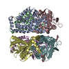
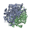
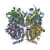
| Deposited unit | 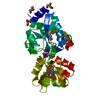
| ||||||||
|---|---|---|---|---|---|---|---|---|---|
| 1 | x 6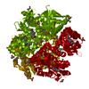
| ||||||||
| Unit cell |
| ||||||||
| Details | Experimentally unknown. It is predicted to be monomeric. |
-Components
-Protein , 1 types, 1 molecules A
| #1: Protein | Mass: 27635.338 Da / Num. of mol.: 1 Source method: isolated from a genetically manipulated source Source: (gene. exp.) Pseudomonas aeruginosa (bacteria) / Strain: PA01 / Gene: PA3475, pheC, Pseudomonas aeruginosa / Plasmid: pMCSG9 / Production host: Escherichia coli (E. coli) / Strain (production host): pPK1037References: UniProt: Q01269, prephenate dehydratase, arogenate dehydratase |
|---|
-Non-polymers , 6 types, 173 molecules 


| #2: Chemical | ChemComp-NI / Nickel Mass: 58.693 Da / Num. of mol.: 1 / Source method: obtained synthetically / Formula: Ni Mass: 58.693 Da / Num. of mol.: 1 / Source method: obtained synthetically / Formula: Ni | ||||||
|---|---|---|---|---|---|---|---|
| #3: Chemical | ChemComp-CL / Chloride Mass: 35.453 Da / Num. of mol.: 1 / Source method: obtained synthetically / Formula: Cl Mass: 35.453 Da / Num. of mol.: 1 / Source method: obtained synthetically / Formula: Cl | ||||||
| #4: Chemical | ChemComp-GOL / Glycerol Mass: 92.094 Da / Num. of mol.: 5 / Source method: obtained synthetically / Formula: C3H8O3 Mass: 92.094 Da / Num. of mol.: 5 / Source method: obtained synthetically / Formula: C3H8O3#5: Chemical | Formic acid Mass: 46.025 Da / Num. of mol.: 2 / Source method: obtained synthetically / Formula: CH2O2 Mass: 46.025 Da / Num. of mol.: 2 / Source method: obtained synthetically / Formula: CH2O2#6: Chemical | ChemComp-EPE / | HEPES Mass: 238.305 Da / Num. of mol.: 1 / Source method: obtained synthetically / Formula: C8H18N2O4S / Comment: pH buffer*YM Mass: 238.305 Da / Num. of mol.: 1 / Source method: obtained synthetically / Formula: C8H18N2O4S / Comment: pH buffer*YM#7: Water | ChemComp-HOH / | WaterMass: 18.015 Da / Num. of mol.: 163 / Source method: isolated from a natural source / Formula: H2O |
-Experimental details
-Experiment
| Experiment | Method: X-RAY DIFFRACTION / Number of used crystals: 1 |
|---|
- Sample preparation
| Crystal | Density Matthews: 2.47 Å3/Da / Density % sol: 50.3 % |
|---|---|
| Crystal grow | Temperature: 289 K / Method: vapor diffusion, sitting drop / pH: 7.5 Details: 0.005M CoCl2 hexahydrate, 0.005M NiCl2 hexahydrate, 0.005M CaCl2 dihydrate, 0.005M MgCl2 hexahydrate, 0.1M HEPES, 12% w/v PEG 3350, pH 7.5, VAPOR DIFFUSION, SITTING DROP, temperature 289K |
-Data collection
| Diffraction | Mean temperature: 100 K |
|---|---|
| Diffraction source | Source: SYNCHROTRON / Site: APS  / Beamline: 19-ID / Wavelength: 0.97948 Å / Beamline: 19-ID / Wavelength: 0.97948 Å |
| Detector | Type: ADSC QUANTUM 315r / Detector: CCD / Date: Aug 11, 2009 / Details: mirror |
| Radiation | Monochromator: Si 111 crystal / Protocol: SINGLE WAVELENGTH / Monochromatic (M) / Laue (L): M / Scattering type: x-ray |
| Radiation wavelength | Wavelength: 0.97948 Å / Relative weight: 1 |
| Reflection | Resolution: 1.659→28.8 Å / Num. all: 32048 / Num. obs: 32048 / % possible obs: 99.1 % / Observed criterion σ(F): 0 / Observed criterion σ(I): 0 / Redundancy: 7.3 % / Rmerge(I) obs: 0.09 / Net I/σ(I): 37.1 |
| Reflection shell | Resolution: 1.66→1.69 Å / Redundancy: 6.9 % / Rmerge(I) obs: 0.795 / Mean I/σ(I) obs: 1.75 / % possible all: 93.9 |
- Processing
| Software |
| ||||||||||||||||||||||||||||||||||||||||||||||||||||||||||||||||||||||||||||||
|---|---|---|---|---|---|---|---|---|---|---|---|---|---|---|---|---|---|---|---|---|---|---|---|---|---|---|---|---|---|---|---|---|---|---|---|---|---|---|---|---|---|---|---|---|---|---|---|---|---|---|---|---|---|---|---|---|---|---|---|---|---|---|---|---|---|---|---|---|---|---|---|---|---|---|---|---|---|---|---|
| Refinement | Method to determine structure: SAD / Resolution: 1.659→28.732 Å / SU ML: 0.21 / σ(F): 1.9 / Phase error: 24.55 / Stereochemistry target values: ML
| ||||||||||||||||||||||||||||||||||||||||||||||||||||||||||||||||||||||||||||||
| Solvent computation | Shrinkage radii: 0.9 Å / VDW probe radii: 1.11 Å / Solvent model: FLAT BULK SOLVENT MODEL / Bsol: 53.051 Å2 / ksol: 0.36 e/Å3 | ||||||||||||||||||||||||||||||||||||||||||||||||||||||||||||||||||||||||||||||
| Refinement step | Cycle: LAST / Resolution: 1.659→28.732 Å
| ||||||||||||||||||||||||||||||||||||||||||||||||||||||||||||||||||||||||||||||
| Refine LS restraints |
| ||||||||||||||||||||||||||||||||||||||||||||||||||||||||||||||||||||||||||||||
| LS refinement shell |
| ||||||||||||||||||||||||||||||||||||||||||||||||||||||||||||||||||||||||||||||
| Refinement TLS params. | Refine-ID: X-RAY DIFFRACTION
| ||||||||||||||||||||||||||||||||||||||||||||||||||||||||||||||||||||||||||||||
| Refinement TLS group | Selection details: chain A resid 124:215 |
