 Movie
Movie Controller

[English] 日本語
 Yorodumi
- PDB-3iv8: N-acetylglucosamine-6-phosphate deacetylase from Vibrio cholerae ... -
+ Open data

- Basic information
| Entry | Database: PDB / ID: 3iv8 | ||||||
|---|---|---|---|---|---|---|---|
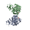
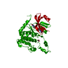
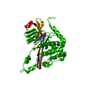
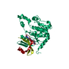
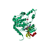
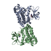
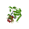
| Title | N-acetylglucosamine-6-phosphate deacetylase from Vibrio cholerae complexed with fructose 6-phosphate | ||||||
 Components Components | N-acetylglucosamine-6-phosphate deacetylase | ||||||
 Keywords Keywords | HYDROLASE / IDP01334 / N-acetylglucosamine-6-phosphate deacetylase / fructose 6-phosphate / Carbohydrate metabolism / Structural Genomics / Center for Structural Genomics of Infectious Diseases / CSGID | ||||||
| Function / homology |  Function and homology informationN-acetylglucosamine-6-phosphate deacetylase / N-acetylgalactosamine-6-phosphate deacetylase activity / N-acetylglucosamine-6-phosphate deacetylase activity / N-acetylglucosamine catabolic process / N-acetylneuraminate catabolic process / protein homotetramerization / carbohydrate metabolic process / metal ion binding Function and homology informationN-acetylglucosamine-6-phosphate deacetylase / N-acetylgalactosamine-6-phosphate deacetylase activity / N-acetylglucosamine-6-phosphate deacetylase activity / N-acetylglucosamine catabolic process / N-acetylneuraminate catabolic process / protein homotetramerization / carbohydrate metabolic process / metal ion bindingSimilarity search - Function | ||||||
| Biological species |   Vibrio cholerae (bacteria) Vibrio cholerae (bacteria) | ||||||
| Method | X-RAY DIFFRACTION / SYNCHROTRON / MOLECULAR REPLACEMENT / Resolution: 2.53 Å | ||||||
 Authors Authors | Osipiuk, J. / Maltseva, N. / Stam, J. / Anderson, W.F. / Joachimiak, A. / Center for Structural Genomics of Infectious Diseases (CSGID) | ||||||
 Citation Citation | Journal: To be Published Title: X-ray crystal structure of N-acetylglucosamine-6-phosphate deacetylase from Vibrio cholerae complexed with fructose 6-phosphate. Authors: Osipiuk, J. / Maltseva, N. / Stam, J. / Anderson, W.F. / Joachimiak, A. | ||||||
| History |
|
- Structure visualization
| Structure viewer | Molecule: MolmilJmol/JSmol |
|---|
- Downloads & links
-Download
| PDBx/mmCIF format | 3iv8.cif.gz | 291.6 KB | Display | PDBx/mmCIF format |
|---|---|---|---|---|
| PDB format | pdb3iv8.ent.gz | 235.3 KB | Display | PDB format |
| PDBx/mmJSON format | 3iv8.json.gz | Tree view | PDBx/mmJSON format | |
| Others |  Other downloads Other downloads |
-Validation report
| Arichive directory | https://data.pdbj.org/pub/pdb/validation_reports/iv/3iv8ftp://data.pdbj.org/pub/pdb/validation_reports/iv/3iv8 | HTTPS FTP |
|---|
-Related structure data
| Related structure data |  3egjS S: Starting model for refinement |
|---|---|
| Similar structure data | |
| Other databases |


-Links
 PDBj
PDBj


- Assembly
| Deposited unit | 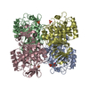
| ||||||||
|---|---|---|---|---|---|---|---|---|---|
| 1 | 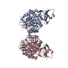
| ||||||||
| 2 | 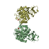
| ||||||||
| Unit cell |
|
-Components
| #1: Protein | / GlcNAc 6-P deacetylase Mass: 41272.000 Da / Num. of mol.: 4 Source method: isolated from a genetically manipulated source Source: (gene. exp.) Vibrio cholerae (bacteria) / Strain: O1 biovar El Tor str. N16961 / Gene: nagA, VC_0994 / Plasmid: pMCSG7 / Production host: Escherichia coli (E. coli) / Strain (production host): BL21(DE3)References: UniProt: O32445, N-acetylglucosamine-6-phosphate deacetylase#2: Sugar | ChemComp-F6P / | Fructose 6-phosphate  Type: D-saccharide, beta linking / Mass: 260.136 Da / Num. of mol.: 1 Type: D-saccharide, beta linking / Mass: 260.136 Da / Num. of mol.: 1Source method: isolated from a genetically manipulated source Formula: C6H13O9P #3: Chemical | ChemComp-NI / Nickel  Mass: 58.693 Da / Num. of mol.: 4 / Source method: obtained synthetically / Formula: Ni Mass: 58.693 Da / Num. of mol.: 4 / Source method: obtained synthetically / Formula: Ni#4: Chemical | Sulfate  Mass: 96.063 Da / Num. of mol.: 3 / Source method: obtained synthetically / Formula: SO4 Mass: 96.063 Da / Num. of mol.: 3 / Source method: obtained synthetically / Formula: SO4#5: Water | ChemComp-HOH / | Water Mass: 18.015 Da / Num. of mol.: 332 / Source method: isolated from a natural source / Formula: H2O Mass: 18.015 Da / Num. of mol.: 332 / Source method: isolated from a natural source / Formula: H2O |
|---|
-Experimental details
-Experiment
| Experiment | Method: X-RAY DIFFRACTION / Number of used crystals: 1 |
|---|
- Sample preparation
| Crystal | Density Matthews: 2.42 Å3/Da / Density % sol: 49.22 % |
|---|---|
| Crystal grow | Temperature: 289 K / Method: vapor diffusion, sitting drop / pH: 7.5 Details: 0.2 M di-sodium hydrogen phosphate, 20% PEG-3350, 5 mM fructose 6-phosphate, pH 7.5, VAPOR DIFFUSION, SITTING DROP, temperature 289K |
-Data collection
| Diffraction | Mean temperature: 100 K |
|---|---|
| Diffraction source | Source: SYNCHROTRON / Site: APS  / Beamline: 19-BM / Wavelength: 0.9792 Å / Beamline: 19-BM / Wavelength: 0.9792 Å |
| Detector | Type: SBC-3 / Detector: CCD / Date: Oct 2, 2008 |
| Radiation | Monochromator: double crystal monochromator / Protocol: SINGLE WAVELENGTH / Monochromatic (M) / Laue (L): M / Scattering type: x-ray |
| Radiation wavelength | Wavelength: 0.9792 Å / Relative weight: 1 |
| Reflection | Resolution: 2.53→35.2 Å / Num. all: 54289 / Num. obs: 54289 / % possible obs: 99.9 % / Observed criterion σ(F): 0 / Observed criterion σ(I): 0 / Redundancy: 6.8 % / Biso Wilson estimate: 48.1 Å2 / Rmerge(I) obs: 0.111 / Χ2: 1.074 / Net I/σ(I): 6.7 |
| Reflection shell | Resolution: 2.53→2.57 Å / Redundancy: 5.3 % / Rmerge(I) obs: 0.68 / Mean I/σ(I) obs: 2.09 / Num. unique all: 2658 / Χ2: 0.827 / % possible all: 100 |
- Processing
| Software |
| |||||||||||||||||||||||||||||||||||||||||||||||||||||||||||||||||||||||||||||||||||||||||||||||||||||||||||||||||||||||||||||
|---|---|---|---|---|---|---|---|---|---|---|---|---|---|---|---|---|---|---|---|---|---|---|---|---|---|---|---|---|---|---|---|---|---|---|---|---|---|---|---|---|---|---|---|---|---|---|---|---|---|---|---|---|---|---|---|---|---|---|---|---|---|---|---|---|---|---|---|---|---|---|---|---|---|---|---|---|---|---|---|---|---|---|---|---|---|---|---|---|---|---|---|---|---|---|---|---|---|---|---|---|---|---|---|---|---|---|---|---|---|---|---|---|---|---|---|---|---|---|---|---|---|---|---|---|---|---|
| Refinement | Method to determine structure: MOLECULAR REPLACEMENT Starting model: pdb entry 3EGJ Resolution: 2.53→35.18 Å / Cor.coef. Fo:Fc: 0.957 / Cor.coef. Fo:Fc free: 0.921 / Occupancy max: 1 / Occupancy min: 0.4 / SU B: 24.772 / SU ML: 0.238 / TLS residual ADP flag: LIKELY RESIDUAL / Cross valid method: THROUGHOUT / σ(F): 0 / σ(I): 0 / ESU R: 0.76 / ESU R Free: 0.311 / Stereochemistry target values: MAXIMUM LIKELIHOOD / Details: HYDROGENS HAVE BEEN ADDED IN THE RIDING POSITIONS
| |||||||||||||||||||||||||||||||||||||||||||||||||||||||||||||||||||||||||||||||||||||||||||||||||||||||||||||||||||||||||||||
| Solvent computation | Ion probe radii: 0.8 Å / Shrinkage radii: 0.8 Å / VDW probe radii: 1.2 Å / Solvent model: MASK | |||||||||||||||||||||||||||||||||||||||||||||||||||||||||||||||||||||||||||||||||||||||||||||||||||||||||||||||||||||||||||||
| Displacement parameters | Biso max: 76.85 Å2 / Biso mean: 34.533 Å2 / Biso min: 10.04 Å2
| |||||||||||||||||||||||||||||||||||||||||||||||||||||||||||||||||||||||||||||||||||||||||||||||||||||||||||||||||||||||||||||
| Refinement step | Cycle: LAST / Resolution: 2.53→35.18 Å
| |||||||||||||||||||||||||||||||||||||||||||||||||||||||||||||||||||||||||||||||||||||||||||||||||||||||||||||||||||||||||||||
| Refine LS restraints |
| |||||||||||||||||||||||||||||||||||||||||||||||||||||||||||||||||||||||||||||||||||||||||||||||||||||||||||||||||||||||||||||
| LS refinement shell | Resolution: 2.53→2.593 Å / Total num. of bins used: 20
| |||||||||||||||||||||||||||||||||||||||||||||||||||||||||||||||||||||||||||||||||||||||||||||||||||||||||||||||||||||||||||||
| Refinement TLS params. | Method: refined / Refine-ID: X-RAY DIFFRACTION
| |||||||||||||||||||||||||||||||||||||||||||||||||||||||||||||||||||||||||||||||||||||||||||||||||||||||||||||||||||||||||||||
| Refinement TLS group |
|
