 Movie
Movie Controller

[English] 日本語
 Yorodumi
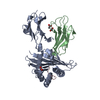
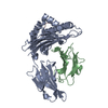
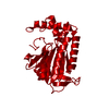
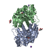
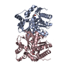
- PDB-3isv: Crystal structure of glutamate racemase from Listeria monocytogen... -
+ Open data

- Basic information
| Entry | Database: PDB / ID: 3isv | ||||||
|---|---|---|---|---|---|---|---|
| Title | Crystal structure of glutamate racemase from Listeria monocytogenes in complex with acetate ion | ||||||
 Components Components | Glutamate racemase | ||||||
 Keywords Keywords | ISOMERASE / glutamate racemase / structural genomics / Cell wall biogenesis/degradation / Peptidoglycan synthesis / Center for Structural Genomics of Infectious Diseases / CSGID / Cell shape | ||||||
| Function / homology |  Function and homology informationglutamate racemase / glutamate racemase activity / peptidoglycan biosynthetic process / cell wall organization / regulation of cell shape Function and homology informationglutamate racemase / glutamate racemase activity / peptidoglycan biosynthetic process / cell wall organization / regulation of cell shapeSimilarity search - Function | ||||||
| Biological species |  Listeria monocytogenes (bacteria) Listeria monocytogenes (bacteria) | ||||||
| Method | X-RAY DIFFRACTION / SYNCHROTRON / MOLECULAR REPLACEMENT / Resolution: 1.85 Å | ||||||
 Authors Authors | Majorek, K.A. / Chruszcz, M. / Skarina, T. / Onopriyenko, O. / Stam, J. / Anderson, W.F. / Savchenko, A. / Bujnicki, J.M. / Minor, W. / Center for Structural Genomics of Infectious Diseases (CSGID) | ||||||
 Citation Citation | Journal: To be Published Title: Crystal structure of glutamate racemase from Listeria monocytogenes in complex with acetate ion Authors: Majorek, K.A. / Chruszcz, M. / Skarina, T. / Onopriyenko, O. / Stam, J. / Anderson, W.F. / Savchenko, A. / Bujnicki, J.M. / Minor, W. / Center for Structural Genomics of Infectious Diseases (CSGID) | ||||||
| History |
|
- Structure visualization
| Structure viewer | Molecule: MolmilJmol/JSmol |
|---|
- Downloads & links
-Download
| PDBx/mmCIF format | 3isv.cif.gz | 126.1 KB | Display | PDBx/mmCIF format |
|---|---|---|---|---|
| PDB format | pdb3isv.ent.gz | 97.5 KB | Display | PDB format |
| PDBx/mmJSON format | 3isv.json.gz | Tree view | PDBx/mmJSON format | |
| Others |  Other downloads Other downloads |
-Validation report
| Arichive directory | https://data.pdbj.org/pub/pdb/validation_reports/is/3isvftp://data.pdbj.org/pub/pdb/validation_reports/is/3isv | HTTPS FTP |
|---|
-Related structure data
| Related structure data |  3hfrS S: Starting model for refinement |
|---|---|
| Similar structure data | |
| Other databases |


-Links
 PDBj
PDBj- Assembly
| Deposited unit | 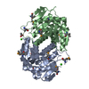
| ||||||||
|---|---|---|---|---|---|---|---|---|---|
| 1 |
| ||||||||
| 2 | 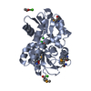
| ||||||||
| 3 | 
| ||||||||
| Unit cell |
| ||||||||
| Components on special symmetry positions |
| ||||||||
| Details | dimer |
-Components
| #1: Protein | Mass: 29795.270 Da / Num. of mol.: 2 Source method: isolated from a genetically manipulated source Source: (gene. exp.) Listeria monocytogenes (bacteria) / Strain: EGD-E / Gene: lmo1237, murI, racE / Plasmid: pMCSG7 / Production host: Escherichia coli (E. coli) / Strain (production host): BL21 DE3 star magic / References: UniProt: Q8Y7N7, glutamate racemase#2: Chemical | Acetate  Mass: 59.044 Da / Num. of mol.: 2 / Source method: obtained synthetically / Formula: C2H3O2 Mass: 59.044 Da / Num. of mol.: 2 / Source method: obtained synthetically / Formula: C2H3O2#3: Chemical |   Mass: 40.078 Da / Num. of mol.: 2 / Source method: obtained synthetically / Formula: Ca Mass: 40.078 Da / Num. of mol.: 2 / Source method: obtained synthetically / Formula: Ca#4: Chemical | Chloride  Mass: 35.453 Da / Num. of mol.: 2 / Source method: obtained synthetically / Formula: Cl Mass: 35.453 Da / Num. of mol.: 2 / Source method: obtained synthetically / Formula: Cl#5: Water | ChemComp-HOH / | Water Mass: 18.015 Da / Num. of mol.: 423 / Source method: isolated from a natural source / Formula: H2O Mass: 18.015 Da / Num. of mol.: 423 / Source method: isolated from a natural source / Formula: H2O |
|---|
-Experimental details
-Experiment
| Experiment | Method: X-RAY DIFFRACTION / Number of used crystals: 1 |
|---|
- Sample preparation
| Crystal | Density Matthews: 2.42 Å3/Da / Density % sol: 49.13 % |
|---|---|
| Crystal grow | Temperature: 293 K / Method: vapor diffusion, hanging drop / pH: 7 Details: PEG8K 18%, CaAcetate 0.2M, NaCacod 0.1m, pH 6.5, 10mM L-Glutamic acid, VAPOR DIFFUSION, HANGING DROP, temperature 293K |
-Data collection
| Diffraction | Mean temperature: 100 K |
|---|---|
| Diffraction source | Source: SYNCHROTRON / Site: APS  / Beamline: 19-ID / Wavelength: 0.97934 Å / Beamline: 19-ID / Wavelength: 0.97934 Å |
| Detector | Type: ADSC QUANTUM 315r / Detector: CCD / Date: Jun 14, 2009 / Details: mirrors |
| Radiation | Monochromator: Si 111 channel / Protocol: SINGLE WAVELENGTH / Monochromatic (M) / Laue (L): M / Scattering type: x-ray |
| Radiation wavelength | Wavelength: 0.97934 Å / Relative weight: 1 |
| Reflection | Resolution: 1.85→50 Å / Num. all: 51135 / Num. obs: 51135 / % possible obs: 99.4 % / Observed criterion σ(F): 0 / Observed criterion σ(I): -3 / Redundancy: 3.9 % / Rmerge(I) obs: 0.084 / Rsym value: 0.084 / Net I/σ(I): 23.3 |
| Reflection shell | Resolution: 1.85→1.88 Å / Redundancy: 4 % / Rmerge(I) obs: 0.621 / Mean I/σ(I) obs: 2 / Rsym value: 0.621 / % possible all: 100 |
- Processing
| Software |
| ||||||||||||||||||||||||||||||||||||||||||||||||||||||||||||||||||||||||||||||||||||||||||||||||||||||||||||||||||||||||||||||||||||||||||||||||||||||||||||||||||||||||||
|---|---|---|---|---|---|---|---|---|---|---|---|---|---|---|---|---|---|---|---|---|---|---|---|---|---|---|---|---|---|---|---|---|---|---|---|---|---|---|---|---|---|---|---|---|---|---|---|---|---|---|---|---|---|---|---|---|---|---|---|---|---|---|---|---|---|---|---|---|---|---|---|---|---|---|---|---|---|---|---|---|---|---|---|---|---|---|---|---|---|---|---|---|---|---|---|---|---|---|---|---|---|---|---|---|---|---|---|---|---|---|---|---|---|---|---|---|---|---|---|---|---|---|---|---|---|---|---|---|---|---|---|---|---|---|---|---|---|---|---|---|---|---|---|---|---|---|---|---|---|---|---|---|---|---|---|---|---|---|---|---|---|---|---|---|---|---|---|---|---|---|---|
| Refinement | Method to determine structure: MOLECULAR REPLACEMENT Starting model: 3hfr Resolution: 1.85→50 Å / Cor.coef. Fo:Fc: 0.964 / Cor.coef. Fo:Fc free: 0.938 / SU B: 6.507 / SU ML: 0.089 / TLS residual ADP flag: LIKELY RESIDUAL / Cross valid method: THROUGHOUT / ESU R: 0.126 / ESU R Free: 0.13 / Stereochemistry target values: MAXIMUM LIKELIHOOD / Details: HYDROGENS HAVE BEEN ADDED IN THE RIDING POSITIONS
| ||||||||||||||||||||||||||||||||||||||||||||||||||||||||||||||||||||||||||||||||||||||||||||||||||||||||||||||||||||||||||||||||||||||||||||||||||||||||||||||||||||||||||
| Solvent computation | Ion probe radii: 0.8 Å / Shrinkage radii: 0.8 Å / VDW probe radii: 1.2 Å / Solvent model: MASK | ||||||||||||||||||||||||||||||||||||||||||||||||||||||||||||||||||||||||||||||||||||||||||||||||||||||||||||||||||||||||||||||||||||||||||||||||||||||||||||||||||||||||||
| Displacement parameters | Biso mean: 12.287 Å2
| ||||||||||||||||||||||||||||||||||||||||||||||||||||||||||||||||||||||||||||||||||||||||||||||||||||||||||||||||||||||||||||||||||||||||||||||||||||||||||||||||||||||||||
| Refinement step | Cycle: LAST / Resolution: 1.85→50 Å
| ||||||||||||||||||||||||||||||||||||||||||||||||||||||||||||||||||||||||||||||||||||||||||||||||||||||||||||||||||||||||||||||||||||||||||||||||||||||||||||||||||||||||||
| Refine LS restraints |
| ||||||||||||||||||||||||||||||||||||||||||||||||||||||||||||||||||||||||||||||||||||||||||||||||||||||||||||||||||||||||||||||||||||||||||||||||||||||||||||||||||||||||||
| LS refinement shell | Resolution: 1.85→1.898 Å / Total num. of bins used: 20
| ||||||||||||||||||||||||||||||||||||||||||||||||||||||||||||||||||||||||||||||||||||||||||||||||||||||||||||||||||||||||||||||||||||||||||||||||||||||||||||||||||||||||||
| Refinement TLS params. | Method: refined / Refine-ID: X-RAY DIFFRACTION
| ||||||||||||||||||||||||||||||||||||||||||||||||||||||||||||||||||||||||||||||||||||||||||||||||||||||||||||||||||||||||||||||||||||||||||||||||||||||||||||||||||||||||||
| Refinement TLS group |
|
