 Movie
Movie Controller


+ Open data

- Basic information
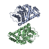
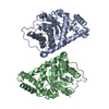
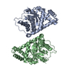
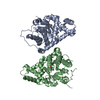
| Entry | Database: PDB / ID: 3cwn | ||||||
|---|---|---|---|---|---|---|---|
| Title | Escherichia coli transaldolase b mutant f178y | ||||||
 Components Components | Transaldolase B | ||||||
 Keywords Keywords | TRANSFERASE / transaldolase / aldolase / directed evolution / Cytoplasm / Pentose shunt | ||||||
| Function / homology |  Function and homology information Function and homology informationtransketolase or transaldolase activity / transaldolase / transaldolase activity / pentose-phosphate shunt, non-oxidative branch / pentose-phosphate shunt / carbohydrate metabolic process / membrane / cytosol Similarity search - Function | ||||||
| Biological species |  | ||||||
| Method |  X-RAY DIFFRACTION / SYNCHROTRON / MOLECULAR REPLACEMENT / Resolution: 1.4 Å X-RAY DIFFRACTION / SYNCHROTRON / MOLECULAR REPLACEMENT / Resolution: 1.4 Å | ||||||
 Authors Authors | Sandalova, T. / Schneider, G. / Samland, A. | ||||||
 Citation Citation | Journal: J.Biol.Chem. / Year: 2008 Title: Replacement of a Phenylalanine by a Tyrosine in the Active Site Confers Fructose-6-phosphate Aldolase Activity to the Transaldolase of Escherichia coli and Human Origin. Authors: Schneider, S. / Sandalova, T. / Schneider, G. / Sprenger, G.A. / Samland, A.K. | ||||||
| History |
|
- Structure visualization
| Structure viewer | Molecule: MolmilJmol/JSmol |
|---|
- Downloads & links
-Download
| PDBx/mmCIF format | 3cwn.cif.gz | 276.6 KB | Display | PDBx/mmCIF format |
|---|---|---|---|---|
| PDB format | pdb3cwn.ent.gz | 224.1 KB | Display | PDB format |
| PDBx/mmJSON format | 3cwn.json.gz | Tree view | PDBx/mmJSON format | |
| Others |  Other downloads Other downloads |
-Validation report
| Arichive directory | https://data.pdbj.org/pub/pdb/validation_reports/cw/3cwnftp://data.pdbj.org/pub/pdb/validation_reports/cw/3cwn | HTTPS FTP |
|---|
-Related structure data
| Related structure data |  1onrS S: Starting model for refinement |
|---|---|
| Similar structure data |


-Links
 PDBj
PDBj- Assembly
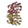
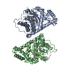
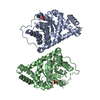
| Deposited unit | 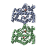
| ||||||||
|---|---|---|---|---|---|---|---|---|---|
| 1 | 
| ||||||||
| 2 | 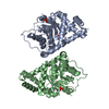
| ||||||||
| Unit cell |
|
-Components
| #1: Protein | Mass: 37476.543 Da / Num. of mol.: 2 / Mutation: F178Y, A247T Source method: isolated from a genetically manipulated source Source: (gene. exp.) References: UniProt: P0A870, UniProt: K0BE10*PLUS, transaldolase #2: Chemical | ChemComp-SO4 /   Mass: 96.063 Da / Num. of mol.: 4 / Source method: obtained synthetically / Formula: SO4 Mass: 96.063 Da / Num. of mol.: 4 / Source method: obtained synthetically / Formula: SO4#3: Water | ChemComp-HOH / |  Mass: 18.015 Da / Num. of mol.: 605 / Source method: isolated from a natural source / Formula: H2O Mass: 18.015 Da / Num. of mol.: 605 / Source method: isolated from a natural source / Formula: H2O |
|---|
-Experimental details
-Experiment
| Experiment | Method: X-RAY DIFFRACTION / Number of used crystals: 1 |
|---|
- Sample preparation
| Crystal | Density Matthews: 2.48 Å3/Da / Density % sol: 50.47 % |
|---|---|
| Crystal grow | Temperature: 290 K / Method: vapor diffusion, hanging drop / pH: 8 Details: 18 % PEG 3350, 0.2 M ammonium sulfate, 25 mM glycylglycine, pH 8.0, VAPOR DIFFUSION, HANGING DROP, temperature 290K |
-Data collection
| Diffraction | Mean temperature: 100 K |
|---|---|
| Diffraction source | Source: SYNCHROTRON / Site: ESRF  / Beamline: ID14-3 / Wavelength: 0.931 Å / Beamline: ID14-3 / Wavelength: 0.931 Å |
| Detector | Type: ADSC QUANTUM 4 / Detector: CCD / Date: Aug 26, 2007 |
| Radiation | Monochromator: diamond / Protocol: SINGLE WAVELENGTH / Monochromatic (M) / Laue (L): M / Scattering type: x-ray |
| Radiation wavelength | Wavelength: 0.931 Å / Relative weight: 1 |
| Reflection | Resolution: 1.4→47 Å / Num. all: 135268 / Num. obs: 135268 / % possible obs: 92 % / Observed criterion σ(F): 0 / Observed criterion σ(I): 0 / Redundancy: 3.7 % / Biso Wilson estimate: 10.2 Å2 / Rmerge(I) obs: 0.037 / Rsym value: 0.037 / Net I/σ(I): 21.4 |
| Reflection shell | Resolution: 1.4→1.48 Å / Redundancy: 2.4 % / Rmerge(I) obs: 0.15 / Mean I/σ(I) obs: 5.7 / Num. unique all: 13203 / Rsym value: 0.15 / % possible all: 62 |
- Processing
| Software |
| ||||||||||||||||||||||||||||||||||||||||||||||||||||||||||||||||||||||||||||||||||||||||||||||||||||||||||||||||||||||||||||||||||||||||||||||||||||||||||||||||||||||||||
|---|---|---|---|---|---|---|---|---|---|---|---|---|---|---|---|---|---|---|---|---|---|---|---|---|---|---|---|---|---|---|---|---|---|---|---|---|---|---|---|---|---|---|---|---|---|---|---|---|---|---|---|---|---|---|---|---|---|---|---|---|---|---|---|---|---|---|---|---|---|---|---|---|---|---|---|---|---|---|---|---|---|---|---|---|---|---|---|---|---|---|---|---|---|---|---|---|---|---|---|---|---|---|---|---|---|---|---|---|---|---|---|---|---|---|---|---|---|---|---|---|---|---|---|---|---|---|---|---|---|---|---|---|---|---|---|---|---|---|---|---|---|---|---|---|---|---|---|---|---|---|---|---|---|---|---|---|---|---|---|---|---|---|---|---|---|---|---|---|---|---|---|
| Refinement | Method to determine structure: MOLECULAR REPLACEMENT Starting model: PDB ENTRY 1ONR Resolution: 1.4→47 Å / Cor.coef. Fo:Fc: 0.972 / Cor.coef. Fo:Fc free: 0.966 / SU B: 1.351 / SU ML: 0.025 / Isotropic thermal model: anisotropic / Cross valid method: THROUGHOUT / σ(F): 0 / σ(I): 0 / ESU R: 0.06 / ESU R Free: 0.053 / Stereochemistry target values: MAXIMUM LIKELIHOOD / Details: HYDROGENS HAVE BEEN ADDED IN THE RIDING POSITIONS
| ||||||||||||||||||||||||||||||||||||||||||||||||||||||||||||||||||||||||||||||||||||||||||||||||||||||||||||||||||||||||||||||||||||||||||||||||||||||||||||||||||||||||||
| Solvent computation | Ion probe radii: 0.8 Å / Shrinkage radii: 0.8 Å / VDW probe radii: 1.4 Å / Solvent model: BABINET MODEL WITH MASK | ||||||||||||||||||||||||||||||||||||||||||||||||||||||||||||||||||||||||||||||||||||||||||||||||||||||||||||||||||||||||||||||||||||||||||||||||||||||||||||||||||||||||||
| Displacement parameters | Biso mean: 9.546 Å2
| ||||||||||||||||||||||||||||||||||||||||||||||||||||||||||||||||||||||||||||||||||||||||||||||||||||||||||||||||||||||||||||||||||||||||||||||||||||||||||||||||||||||||||
| Refinement step | Cycle: LAST / Resolution: 1.4→47 Å
| ||||||||||||||||||||||||||||||||||||||||||||||||||||||||||||||||||||||||||||||||||||||||||||||||||||||||||||||||||||||||||||||||||||||||||||||||||||||||||||||||||||||||||
| Refine LS restraints |
| ||||||||||||||||||||||||||||||||||||||||||||||||||||||||||||||||||||||||||||||||||||||||||||||||||||||||||||||||||||||||||||||||||||||||||||||||||||||||||||||||||||||||||
| LS refinement shell | Resolution: 1.4→1.436 Å / Total num. of bins used: 20
|
