 Movie
Movie Controller


+ Open data

- Basic information
| Entry | Database: PDB / ID: 3cro | ||||||
|---|---|---|---|---|---|---|---|
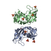
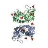
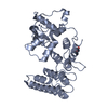
| Title | THE PHAGE 434 CRO/OR1 COMPLEX AT 2.5 ANGSTROMS RESOLUTION | ||||||
 Components Components |
| ||||||
 Keywords Keywords | TRANSCRIPTION/DNA / PROTEIN-DNA COMPLEX / DOUBLE HELIX / TRANSCRIPTION-DNA COMPLEX | ||||||
| Function / homology |  Function and homology information Function and homology information | ||||||
| Biological species |  Phage 434 (virus) Phage 434 (virus) | ||||||
| Method |  X-RAY DIFFRACTION / Resolution: 2.5 Å X-RAY DIFFRACTION / Resolution: 2.5 Å | ||||||
 Authors Authors | Mondragon, A. / Harrison, S.C. | ||||||
 Citation Citation | Journal: J.Mol.Biol. / Year: 1991 Title: The phage 434 Cro/OR1 complex at 2.5 A resolution. Authors: Mondragon, A. / Harrison, S.C. #1: Journal: J.Mol.Biol. / Year: 1989Title: Structure of Phage 434 Cro Protein at 2.35 Angstroms Resolution Authors: Mondragon, A. / Wolberger, C. / Harrison, S.C. #2: Journal: Science / Year: 1988Title: Recognition of a DNA Operator by the Repressor of Phage 434. A View at High Resolution Authors: Aggarwal, A.K. / Rodgers, D.W. / Drottar, M. / Ptashne, M. / Harrison, S.C. #3: Journal: Nature / Year: 1987Title: Structure of the Repressor-Operator Complex of Bacteriophage 434 Authors: Anderson, J.E. / Ptashne, M. / Harrison, S.C. | ||||||
| History |
|
- Structure visualization
| Structure viewer | Molecule: MolmilJmol/JSmol |
|---|
- Downloads & links
-Download
| PDBx/mmCIF format | 3cro.cif.gz | 61.4 KB | Display | PDBx/mmCIF format |
|---|---|---|---|---|
| PDB format | pdb3cro.ent.gz | 42.7 KB | Display | PDB format |
| PDBx/mmJSON format | 3cro.json.gz | Tree view | PDBx/mmJSON format | |
| Others |  Other downloads Other downloads |
-Validation report
| Summary document | 3cro_validation.pdf.gz | 430.5 KB | Display | wwPDB validaton report |
|---|---|---|---|---|
| Full document | 3cro_full_validation.pdf.gz | 449.4 KB | Display | |
| Data in XML | 3cro_validation.xml.gz | 10.9 KB | Display | |
| Data in CIF | 3cro_validation.cif.gz | 14.5 KB | Display | |
| Arichive directory | https://data.pdbj.org/pub/pdb/validation_reports/cr/3croftp://data.pdbj.org/pub/pdb/validation_reports/cr/3cro | HTTPS FTP |
-Related structure data
| Similar structure data |
|---|


-Links
 PDBj
PDBj


- Assembly
| Deposited unit | 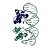
| ||||||||
|---|---|---|---|---|---|---|---|---|---|
| 1 |
| ||||||||
| Unit cell |
| ||||||||
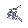
| Details | THE TWO PROTEIN DOMAINS HAVE BEEN ASSIGNED CHAIN INDICATORS *L* AND *R*. THE TWO DNA CHAINS HAVE BEEN ASSIGNED CHAIN INDICATORS *A* AND *B*. THE UNIT CELL CONTAINS TWO COMPLEXES EACH OF WHICH CONSISTS OF A PROTEIN DIMER AND TWO DIFFERENT STRANDS OF DNA. |
-Components
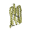
| #1: DNA chain | Mass: 6106.990 Da / Num. of mol.: 1 / Source method: obtained synthetically | ||||
|---|---|---|---|---|---|
| #2: DNA chain | Mass: 6156.027 Da / Num. of mol.: 1 / Source method: obtained synthetically | ||||
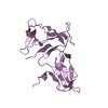
| #3: Protein | Mass: 8076.619 Da / Num. of mol.: 2 / Source method: isolated from a natural source / Source: (natural) Phage 434 (virus) / Genus: Lambda-like viruses / Species: Enterobacteria phage lambda / References: UniProt: P03036#4: Water | ChemComp-HOH / |  Mass: 18.015 Da / Num. of mol.: 25 / Source method: isolated from a natural source / Formula: H2O Mass: 18.015 Da / Num. of mol.: 25 / Source method: isolated from a natural source / Formula: H2OSequence details | THE DNA CHAINS ARE ALIGNED AS FOLLOWS *B* 5(PRIME) T-A-T-A-C-A-A-G-A-A-A-G-T-T-T-G-T-A-C-T *A* ...THE DNA CHAINS ARE ALIGNED AS FOLLOWS *B* 5(PRIME) T-A-T-A-C-A-A-G-A-A-A-G-T-T-T-G-T-A-C-T *A* 3(PRIME) T-A-T-G-T-T-C-T-T-T-C-A-A-A-C-A-T-G-A-A THE DNA CHAINS ARE NUMBERED SEQUENTIAL | |
-Experimental details
-Experiment
| Experiment | Method: X-RAY DIFFRACTION |
|---|
- Sample preparation
| Crystal | Density Matthews: 2.36 Å3/Da / Density % sol: 47.95 % | ||||||||||||||||||||||||||||||||||||||||||||||||||||||||
|---|---|---|---|---|---|---|---|---|---|---|---|---|---|---|---|---|---|---|---|---|---|---|---|---|---|---|---|---|---|---|---|---|---|---|---|---|---|---|---|---|---|---|---|---|---|---|---|---|---|---|---|---|---|---|---|---|---|
| Crystal grow | Temperature: 277 K / pH: 6.2 / Details: pH 6.20, temperature 277.00K | ||||||||||||||||||||||||||||||||||||||||||||||||||||||||
| Components of the solutions |
| ||||||||||||||||||||||||||||||||||||||||||||||||||||||||
| Crystal grow | *PLUS Temperature: 4 ℃ / Method: unknown / pH: 6.2 | ||||||||||||||||||||||||||||||||||||||||||||||||||||||||
| Components of the solutions | *PLUS
|
-Data collection
| Diffraction | Mean temperature: 277 K |
|---|---|
| Diffraction source | Source: ROTATING ANODE / Type: ELLIOTT GX-13 |
| Detector | Type: XENTRONICS / Detector: AREA DETECTOR |
| Radiation | Protocol: SINGLE WAVELENGTH / Monochromatic (M) / Laue (L): M / Scattering type: x-ray |
| Radiation wavelength | Relative weight: 1 |
| Reflection | Highest resolution: 2.5 Å / Num. obs: 7232 |
| Reflection | *PLUS Highest resolution: 2.5 Å / Rmerge(I) obs: 0.101 |
- Processing
| Software | Name: TNT / Classification: refinement | ||||||||||||||||||||||||||||||
|---|---|---|---|---|---|---|---|---|---|---|---|---|---|---|---|---|---|---|---|---|---|---|---|---|---|---|---|---|---|---|---|
| Refinement | Resolution: 2.5→10 Å Details: DENSITY FOR THE LAST SIX CARBOXY TERMINAL RESIDUES (64 - 70) WAS NOT OBSERVED IN THE MAP AT ANY STAGE OF THE REFINEMENT AND THESE RESIDUES ARE PROBABLY DISORDERED.
| ||||||||||||||||||||||||||||||
| Refinement step | Cycle: LAST / Resolution: 2.5→10 Å
| ||||||||||||||||||||||||||||||
| Refine LS restraints |
| ||||||||||||||||||||||||||||||
| Refinement | *PLUS Highest resolution: 2.5 Å / Lowest resolution: 10 Å / Rfactor obs: 0.22 | ||||||||||||||||||||||||||||||
| Solvent computation | *PLUS | ||||||||||||||||||||||||||||||
| Displacement parameters | *PLUS | ||||||||||||||||||||||||||||||
| Refine LS restraints | *PLUS
|
