 Movie
Movie Controller

[English] 日本語
 Yorodumi
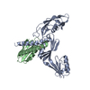
- PDB-2z7e: Crystal structure of Aquifex aeolicus IscU with bound [2Fe-2S] cluster -
+ Open data

- Basic information
| Entry | Database: PDB / ID: 2z7e | ||||||
|---|---|---|---|---|---|---|---|
| Title | Crystal structure of Aquifex aeolicus IscU with bound [2Fe-2S] cluster | ||||||
 Components Components | NifU-like protein | ||||||
 Keywords Keywords | BIOSYNTHETIC PROTEIN / iron-sulfur cluster / iron / biosynthesis / [2Fe-2S] / ISC / IscU / NifU / asymmetric trimer / three conserved Cys | ||||||
| Function / homology |  Function and homology information Function and homology informationiron-sulfur cluster assembly / ferrous iron binding / 2 iron, 2 sulfur cluster binding / intracellular iron ion homeostasis / cytoplasm Similarity search - Function | ||||||
| Biological species |   Aquifex aeolicus (bacteria) Aquifex aeolicus (bacteria) | ||||||
| Method |  X-RAY DIFFRACTION / SYNCHROTRON / MAD / Resolution: 2.3 Å X-RAY DIFFRACTION / SYNCHROTRON / MAD / Resolution: 2.3 Å | ||||||
 Authors Authors | Shimomura, Y. / Wada, K. / Takahashi, Y. / Fukuyama, K. | ||||||
 Citation Citation | Journal: J.Mol.Biol. / Year: 2008 Title: The asymmetric trimeric architecture of [2Fe-2S] IscU: implications for its scaffolding during iron-sulfur cluster biosynthesis Authors: Shimomura, Y. / Wada, K. / Fukuyama, K. / Takahashi, Y. | ||||||
| History |
|
- Structure visualization
| Structure viewer | Molecule: MolmilJmol/JSmol |
|---|
- Downloads & links
-Download
| PDBx/mmCIF format | 2z7e.cif.gz | 93.6 KB | Display | PDBx/mmCIF format |
|---|---|---|---|---|
| PDB format | pdb2z7e.ent.gz | 72.1 KB | Display | PDB format |
| PDBx/mmJSON format | 2z7e.json.gz | Tree view | PDBx/mmJSON format | |
| Others |  Other downloads Other downloads |
-Validation report
| Arichive directory | https://data.pdbj.org/pub/pdb/validation_reports/z7/2z7eftp://data.pdbj.org/pub/pdb/validation_reports/z7/2z7e | HTTPS FTP |
|---|
-Related structure data
| Similar structure data |
|---|


-Links
 PDBj
PDBj


- Assembly
| Deposited unit | 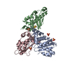
| ||||||||
|---|---|---|---|---|---|---|---|---|---|
| 1 |
| ||||||||
| 2 | 
| ||||||||
| Unit cell |
| ||||||||
| Components on special symmetry positions |
| ||||||||
| Details | The biological assembly is a trimer which consists of the three protomers in the asymmetric unit. |
-Components
| #1: Protein | Mass: 17503.795 Da / Num. of mol.: 3 / Mutation: D38A Source method: isolated from a genetically manipulated source Source: (gene. exp.) Aquifex aeolicus (bacteria) / Strain: VF5 / Gene: AQ_896 / Plasmid: pET21a(+) / Production host: #2: Chemical | ChemComp-SO4 /   Mass: 96.063 Da / Num. of mol.: 4 / Source method: obtained synthetically / Formula: SO4 Mass: 96.063 Da / Num. of mol.: 4 / Source method: obtained synthetically / Formula: SO4#3: Chemical | ChemComp-FES / |   Mass: 175.820 Da / Num. of mol.: 1 / Source method: obtained synthetically / Formula: Fe2S2 Mass: 175.820 Da / Num. of mol.: 1 / Source method: obtained synthetically / Formula: Fe2S2#4: Water | ChemComp-HOH / |  Mass: 18.015 Da / Num. of mol.: 162 / Source method: isolated from a natural source / Formula: H2O Mass: 18.015 Da / Num. of mol.: 162 / Source method: isolated from a natural source / Formula: H2OHas protein modification | Y | |
|---|
-Experimental details
-Experiment
| Experiment | Method: X-RAY DIFFRACTION / Number of used crystals: 1 |
|---|
- Sample preparation
| Crystal | Density Matthews: 2.63 Å3/Da / Density % sol: 53.24 % |
|---|---|
| Crystal grow | Temperature: 293 K / Method: vapor diffusion, hanging drop Details: 1.6M ammonium sulfate, 0.5M lithium chloride, VAPOR DIFFUSION, HANGING DROP, temperature 293K |
-Data collection
| Diffraction | Mean temperature: 100 K | |||||||||||||||
|---|---|---|---|---|---|---|---|---|---|---|---|---|---|---|---|---|
| Diffraction source | Source: SYNCHROTRON / Site: SPring-8  / Beamline: BL41XU / Wavelength: 1.7407, 1.7423, 1.6947, 1.5000 / Beamline: BL41XU / Wavelength: 1.7407, 1.7423, 1.6947, 1.5000 | |||||||||||||||
| Detector | Type: ADSC QUANTUM 315 / Detector: CCD / Date: Mar 26, 2006 | |||||||||||||||
| Radiation | Protocol: MAD / Monochromatic (M) / Laue (L): M / Scattering type: x-ray | |||||||||||||||
| Radiation wavelength |
| |||||||||||||||
| Reflection | Resolution: 2.3→47.25 Å / Num. obs: 24440 / % possible obs: 96.5 % / Biso Wilson estimate: 28 Å2 |
- Processing
| Software |
| ||||||||||||||||||||||||||||||||||||||||||||||||||||||||||||||||||||||||||||||||
|---|---|---|---|---|---|---|---|---|---|---|---|---|---|---|---|---|---|---|---|---|---|---|---|---|---|---|---|---|---|---|---|---|---|---|---|---|---|---|---|---|---|---|---|---|---|---|---|---|---|---|---|---|---|---|---|---|---|---|---|---|---|---|---|---|---|---|---|---|---|---|---|---|---|---|---|---|---|---|---|---|---|
| Refinement | Method to determine structure: MAD / Resolution: 2.3→47.25 Å / Rfactor Rfree error: 0.007 / Data cutoff high absF: 1635058.45 / Data cutoff low absF: 0 / Isotropic thermal model: RESTRAINED / Cross valid method: THROUGHOUT / σ(F): 0
| ||||||||||||||||||||||||||||||||||||||||||||||||||||||||||||||||||||||||||||||||
| Solvent computation | Solvent model: FLAT MODEL / Bsol: 74.561 Å2 / ksol: 0.38913 e/Å3 | ||||||||||||||||||||||||||||||||||||||||||||||||||||||||||||||||||||||||||||||||
| Displacement parameters | Biso mean: 49.9 Å2
| ||||||||||||||||||||||||||||||||||||||||||||||||||||||||||||||||||||||||||||||||
| Refine analyze |
| ||||||||||||||||||||||||||||||||||||||||||||||||||||||||||||||||||||||||||||||||
| Refinement step | Cycle: LAST / Resolution: 2.3→47.25 Å
| ||||||||||||||||||||||||||||||||||||||||||||||||||||||||||||||||||||||||||||||||
| Refine LS restraints |
| ||||||||||||||||||||||||||||||||||||||||||||||||||||||||||||||||||||||||||||||||
| LS refinement shell | Resolution: 2.3→2.44 Å / Rfactor Rfree error: 0.022 / Total num. of bins used: 6
| ||||||||||||||||||||||||||||||||||||||||||||||||||||||||||||||||||||||||||||||||
| Xplor file |
|
