 Movie
Movie Controller

[English] 日本語
 Yorodumi
- PDB-2yyj: Crystal structure of the oxygenase component (HpaB) of 4-hydroxyp... -
+ Open data

- Basic information
| Entry | Database: PDB / ID: 2yyj | ||||||
|---|---|---|---|---|---|---|---|
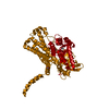
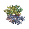
| Title | Crystal structure of the oxygenase component (HpaB) of 4-hydroxyphenylacetate 3-monooxygenase complexed with FAD and 4-hydroxyphenylacetate | ||||||
 Components Components | 4-hydroxyphenylacetate-3-hydroxylase | ||||||
 Keywords Keywords |  OXIDOREDUCTASE / Structurome / RIKEN SPring-8 Center / oxygnase component / 4-hydroxyphenylacetate 3-monooxygenase / two-component flavin diffusible monooxygenase / FAD and a substrate complex OXIDOREDUCTASE / Structurome / RIKEN SPring-8 Center / oxygnase component / 4-hydroxyphenylacetate 3-monooxygenase / two-component flavin diffusible monooxygenase / FAD and a substrate complex | ||||||
| Function / homology |  Function and homology information4-hydroxyphenylacetate 3-monooxygenase / 4-hydroxyphenylacetate 3-monooxygenase activity / phenylacetate catabolic process / oxidoreductase activity, acting on the CH-CH group of donors / flavin adenine dinucleotide binding Function and homology information4-hydroxyphenylacetate 3-monooxygenase / 4-hydroxyphenylacetate 3-monooxygenase activity / phenylacetate catabolic process / oxidoreductase activity, acting on the CH-CH group of donors / flavin adenine dinucleotide bindingSimilarity search - Function | ||||||
| Biological species |   Thermus thermophilus (bacteria) Thermus thermophilus (bacteria) | ||||||
| Method | X-RAY DIFFRACTION / SYNCHROTRON / MOLECULAR REPLACEMENT / Resolution: 1.66 Å | ||||||
 Authors Authors | Kim, S.-H. / Hisano, T. / Takeda, K. / Iwasaki, W. / Ebihara, A. / Miki, K. | ||||||
 Citation Citation | Journal: J.Biol.Chem. / Year: 2007 Title: Crystal Structure of the Oxygenase Component (HpaB) of the 4-Hydroxyphenylacetate 3-Monooxygenase from Thermus thermophilus HB8 Authors: Kim, S.-H. / Hisano, T. / Takeda, K. / Iwasaki, W. / Ebihara, A. / Miki, K. #1: Journal: Acta Crystallogr.,Sect.F / Year: 2007 Title: Crystallization and preliminary X-ray analysis of the oxygenase component (HpaB) of 4-hydroxyphenylacetate 3-monooxygenase from Thermus thermophilus HB8 Authors: Kim, S.-H. / Miyatake, H. / Hisano, T. / Iwasaki, W. / Ebihara, A. / Miki, K. | ||||||
| History |
|
- Structure visualization
| Structure viewer | Molecule: MolmilJmol/JSmol |
|---|
- Downloads & links
-Download
| PDBx/mmCIF format | 2yyj.cif.gz | 122.9 KB | Display | PDBx/mmCIF format |
|---|---|---|---|---|
| PDB format | pdb2yyj.ent.gz | 92.6 KB | Display | PDB format |
| PDBx/mmJSON format | 2yyj.json.gz | Tree view | PDBx/mmJSON format | |
| Others |  Other downloads Other downloads |
-Validation report
| Arichive directory | https://data.pdbj.org/pub/pdb/validation_reports/yy/2yyjftp://data.pdbj.org/pub/pdb/validation_reports/yy/2yyj | HTTPS FTP |
|---|
-Related structure data
| Related structure data |  2yygC  2yyiC  2yykSC  2yylC  2yymC C: citing same article ( S: Starting model for refinement |
|---|---|
| Similar structure data |


-Links
 PDBj
PDBj- Assembly
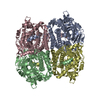
| Deposited unit | 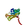
| |||||||||
|---|---|---|---|---|---|---|---|---|---|---|
| 1 | 
| |||||||||
| Unit cell |
| |||||||||
| Components on special symmetry positions |
|
-Components
| #1: Protein | Mass: 54369.043 Da / Num. of mol.: 1 Source method: isolated from a genetically manipulated source Source: (gene. exp.) Thermus thermophilus (bacteria) / Strain: HB8 / Plasmid: pET11a / Species (production host): Escherichia coli / Production host: Escherichia coli BL21(DE3) (bacteria) / Strain (production host): BL21(DE3) / References: UniProt: Q5SJP8, EC: 1.14.13.3 | ||||||
|---|---|---|---|---|---|---|---|
| #2: Chemical | ChemComp-SO4 / Sulfate  Mass: 96.063 Da / Num. of mol.: 6 / Source method: obtained synthetically / Formula: SO4 Mass: 96.063 Da / Num. of mol.: 6 / Source method: obtained synthetically / Formula: SO4#3: Chemical | ChemComp-FAD / | Flavin adenine dinucleotide  Mass: 785.550 Da / Num. of mol.: 1 / Source method: obtained synthetically / Formula: C27H33N9O15P2 / Comment: FAD*YM Mass: 785.550 Da / Num. of mol.: 1 / Source method: obtained synthetically / Formula: C27H33N9O15P2 / Comment: FAD*YM#4: Chemical | 4-Hydroxyphenylacetic acid  Mass: 152.147 Da / Num. of mol.: 2 / Source method: obtained synthetically / Formula: C8H8O3 Mass: 152.147 Da / Num. of mol.: 2 / Source method: obtained synthetically / Formula: C8H8O3#5: Water | ChemComp-HOH / | Water Mass: 18.015 Da / Num. of mol.: 561 / Source method: isolated from a natural source / Formula: H2O Mass: 18.015 Da / Num. of mol.: 561 / Source method: isolated from a natural source / Formula: H2O |
-Experimental details
-Experiment
| Experiment | Method: X-RAY DIFFRACTION / Number of used crystals: 1 |
|---|
- Sample preparation
| Crystal | Density Matthews: 2.69 Å3/Da / Density % sol: 54.2 % |
|---|---|
| Crystal grow | Temperature: 293 K / Method: vapor diffusion, hanging drop / pH: 8.5 Details: 1.5M ammonium sulfate, 0.1M Tris-HCl, 25% (v/v) glycerol, pH 8.5, VAPOR DIFFUSION, HANGING DROP, temperature 293K |
-Data collection
| Diffraction | Mean temperature: 100 K |
|---|---|
| Diffraction source | Source: SYNCHROTRON / Site: SPring-8  / Beamline: BL44B2 / Wavelength: 1 Å / Beamline: BL44B2 / Wavelength: 1 Å |
| Detector | Type: ADSC QUANTUM 210 / Detector: CCD / Date: Sep 21, 2005 / Details: mirrors |
| Radiation | Monochromator: Si / Protocol: SINGLE WAVELENGTH / Monochromatic (M) / Laue (L): M / Scattering type: x-ray |
| Radiation wavelength | Wavelength: 1 Å / Relative weight: 1 |
| Reflection | Resolution: 1.66→46.69 Å / Num. all: 68617 / Num. obs: 68617 / Redundancy: 5.9 % / Biso Wilson estimate: 16.542 Å2 / Rmerge(I) obs: 0.059 / Net I/σ(I): 21.4 |
| Reflection shell | Resolution: 1.66→1.72 Å / Redundancy: 4.2 % / Rmerge(I) obs: 0.208 / Num. unique all: 6416 |
- Processing
| Software |
| ||||||||||||||||||||||||||||||||||||||||||
|---|---|---|---|---|---|---|---|---|---|---|---|---|---|---|---|---|---|---|---|---|---|---|---|---|---|---|---|---|---|---|---|---|---|---|---|---|---|---|---|---|---|---|---|
| Refinement | Method to determine structure: MOLECULAR REPLACEMENT Starting model: PDB ENTRY 2YYK Resolution: 1.66→46.69 Å / Isotropic thermal model: Isotropic / Cross valid method: THROUGHOUT / σ(F): 0 / Stereochemistry target values: Engh & Huber
| ||||||||||||||||||||||||||||||||||||||||||
| Displacement parameters | Biso mean: 17.3 Å2
| ||||||||||||||||||||||||||||||||||||||||||
| Refine analyze |
| ||||||||||||||||||||||||||||||||||||||||||
| Refinement step | Cycle: LAST / Resolution: 1.66→46.69 Å
| ||||||||||||||||||||||||||||||||||||||||||
| Refine LS restraints |
| ||||||||||||||||||||||||||||||||||||||||||
| LS refinement shell | Refine-ID: X-RAY DIFFRACTION / Total num. of bins used: 6
|
