 Movie
Movie Controller

[English] 日本語
 Yorodumi
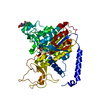
- PDB-2xut: Crystal structure of a proton dependent oligopeptide (POT) family... -
+ Open data

- Basic information
| Entry | Database: PDB / ID: 2xut | ||||||
|---|---|---|---|---|---|---|---|
| Title | Crystal structure of a proton dependent oligopeptide (POT) family transporter. | ||||||
 Components Components | PROTON/PEPTIDE SYMPORTER FAMILY PROTEIN | ||||||
 Keywords Keywords | TRANSPORT PROTEIN / MEMBRANE PROTEIN / MAJOR FACILITATOR SUPERFAMILY TRANSPORTER / PROTON COUPLED PEPTIDE TRANSPORT | ||||||
| Function / homology |  Function and homology information Function and homology informationoligopeptide transport / peptide transmembrane transporter activity / transmembrane transporter activity / transmembrane transport / membrane / plasma membrane Similarity search - Function | ||||||
| Biological species |  SHEWANELLA ONEIDENSIS (bacteria) SHEWANELLA ONEIDENSIS (bacteria) | ||||||
| Method |  X-RAY DIFFRACTION / SYNCHROTRON / MIRAS / Resolution: 3.62 Å X-RAY DIFFRACTION / SYNCHROTRON / MIRAS / Resolution: 3.62 Å | ||||||
 Authors Authors | Newstead, S. / Drew, D. / Cameron, A.D. / Postis, V.L. / Xia, X. / Fowler, P.W. / Ingram, J.C. / Carpenter, E.P. / Sansom, M.S.P. / McPherson, M.J. ...Newstead, S. / Drew, D. / Cameron, A.D. / Postis, V.L. / Xia, X. / Fowler, P.W. / Ingram, J.C. / Carpenter, E.P. / Sansom, M.S.P. / McPherson, M.J. / Baldwin, S.A. / Iwata, S. | ||||||
 Citation Citation | Journal: Embo J. / Year: 2011 Title: Crystal Structure of a Prokaryotic Homologue of the Mammalian Oligopeptide-Proton Symporters, Pept1 and Pept2. Authors: Newstead, S. / Drew, D. / Cameron, A.D. / Postis, V.L. / Xia, X. / Fowler, P.W. / Ingram, J.C. / Carpenter, E.P. / Sansom, M.S.P. / McPherson, M.J. / Baldwin, S.A. / Iwata, S. | ||||||
| History |
|
- Structure visualization
| Structure viewer | Molecule: MolmilJmol/JSmol |
|---|
- Downloads & links
-Download
| PDBx/mmCIF format | 2xut.cif.gz | 466 KB | Display | PDBx/mmCIF format |
|---|---|---|---|---|
| PDB format | pdb2xut.ent.gz | 392.1 KB | Display | PDB format |
| PDBx/mmJSON format | 2xut.json.gz | Tree view | PDBx/mmJSON format | |
| Others |  Other downloads Other downloads |
-Validation report
| Arichive directory | https://data.pdbj.org/pub/pdb/validation_reports/xu/2xutftp://data.pdbj.org/pub/pdb/validation_reports/xu/2xut | HTTPS FTP |
|---|
-Related structure data
| Similar structure data |
|---|


-Links
 PDBj
PDBj


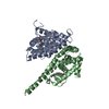
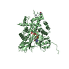
- Assembly
| Deposited unit | 
| ||||||||||||
|---|---|---|---|---|---|---|---|---|---|---|---|---|---|
| 1 | 
| ||||||||||||
| 2 | 
| ||||||||||||
| 3 | 
| ||||||||||||
| Unit cell |
| ||||||||||||
| Noncrystallographic symmetry (NCS) | NCS oper:
|
-Components
| #1: Protein | Mass: 57684.824 Da / Num. of mol.: 3 / Mutation: YES Source method: isolated from a genetically manipulated source Source: (gene. exp.) SHEWANELLA ONEIDENSIS (bacteria) / Strain: MR-1 / Plasmid: PWALDO-GFPE / Production host: Compound details | ENGINEERED RESIDUE IN CHAIN A, THR 2 TO ASN ENGINEERED RESIDUE IN CHAIN A, THR 3 TO SER ENGINEERED ...ENGINEERED | |
|---|
-Experimental details
-Experiment
| Experiment | Method: X-RAY DIFFRACTION / Number of used crystals: 1 |
|---|
- Sample preparation
| Crystal | Density Matthews: 6.49 Å3/Da / Density % sol: 80 % Description: REFINEMENT WAS CARRIED OUT USING ANISOTROPIC TRUNCATION OF THE OBSERVED STRUCTURE FACTORS TO 4.3 A ALONG THE A AND B AXES, AND 3.6 ALONG C |
|---|---|
| Crystal grow | pH: 6.5 / Details: 30% PEG 300, 0.1M MES PH 6.50 AND 0.1M NACL |
-Data collection
| Diffraction | Mean temperature: 277 K |
|---|---|
| Diffraction source | Source: SYNCHROTRON / Site: Diamond  / Beamline: I02 / Wavelength: 0.9795 / Beamline: I02 / Wavelength: 0.9795 |
| Detector | Type: ADSC CCD / Detector: CCD / Date: Feb 18, 2009 Details: KIRKPATRICK BAEZ BIMORPH MIRROR PAIR FOR HORIZONTAL AND VERTICAL FOCUSSING |
| Radiation | Monochromator: SI (111) DOUBLE CRYSTAL MONOCHROMATOR / Protocol: SINGLE WAVELENGTH / Monochromatic (M) / Laue (L): M / Scattering type: x-ray |
| Radiation wavelength | Wavelength: 0.9795 Å / Relative weight: 1 |
| Reflection | Resolution: 3.62→35.5 Å / Num. obs: 47021 / % possible obs: 93.8 % / Observed criterion σ(I): 1.1 / Redundancy: 2 % / Biso Wilson estimate: 105.41 Å2 / Rmerge(I) obs: 0.09 / Net I/σ(I): 9 |
| Reflection shell | Resolution: 3.6→3.73 Å / Redundancy: 1.8 % / Rmerge(I) obs: 0.68 / Mean I/σ(I) obs: 1.1 / % possible all: 88.7 |
- Processing
| Software |
| ||||||||||||||||||||||||||||||||||||||||||||||||||||||||||||||||||||||||||||||||||||||||||||||||||||||||||||||||||
|---|---|---|---|---|---|---|---|---|---|---|---|---|---|---|---|---|---|---|---|---|---|---|---|---|---|---|---|---|---|---|---|---|---|---|---|---|---|---|---|---|---|---|---|---|---|---|---|---|---|---|---|---|---|---|---|---|---|---|---|---|---|---|---|---|---|---|---|---|---|---|---|---|---|---|---|---|---|---|---|---|---|---|---|---|---|---|---|---|---|---|---|---|---|---|---|---|---|---|---|---|---|---|---|---|---|---|---|---|---|---|---|---|---|---|---|
| Refinement | Method to determine structure: MIRAS / Resolution: 3.62→35.47 Å / Cross valid method: THROUGHOUT / σ(F): 0 Details: THE MODEL WAS BUILT AND REFINED USING DATA WITH ANISOTROPIC TRUNCATION OF THE OBSERVED STRUCTURE A AND B AXES, WHILST KEEPING THE C AXIS AT 3.6A.
| ||||||||||||||||||||||||||||||||||||||||||||||||||||||||||||||||||||||||||||||||||||||||||||||||||||||||||||||||||
| Displacement parameters | Biso mean: 252.95 Å2
| ||||||||||||||||||||||||||||||||||||||||||||||||||||||||||||||||||||||||||||||||||||||||||||||||||||||||||||||||||
| Refine analyze | Luzzati coordinate error obs: 1.877 Å | ||||||||||||||||||||||||||||||||||||||||||||||||||||||||||||||||||||||||||||||||||||||||||||||||||||||||||||||||||
| Refinement step | Cycle: LAST / Resolution: 3.62→35.47 Å
| ||||||||||||||||||||||||||||||||||||||||||||||||||||||||||||||||||||||||||||||||||||||||||||||||||||||||||||||||||
| Refine LS restraints |
| ||||||||||||||||||||||||||||||||||||||||||||||||||||||||||||||||||||||||||||||||||||||||||||||||||||||||||||||||||
| LS refinement shell | Resolution: 3.62→3.76 Å / Total num. of bins used: 14
| ||||||||||||||||||||||||||||||||||||||||||||||||||||||||||||||||||||||||||||||||||||||||||||||||||||||||||||||||||
| Refinement TLS params. | L33: 16.6309 °2 / Method: refined / Refine-ID: X-RAY DIFFRACTION
| ||||||||||||||||||||||||||||||||||||||||||||||||||||||||||||||||||||||||||||||||||||||||||||||||||||||||||||||||||
| Refinement TLS group |
|
