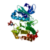
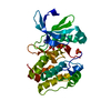
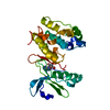
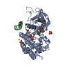
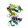
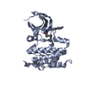
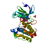
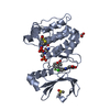
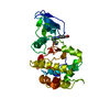
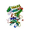
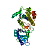
Interaction between PHLDA1 and AURKA / regulation of centrosome cycle / axon hillock / spindle assembly involved in female meiosis I / cilium disassembly / spindle pole centrosome / chromosome passenger complex / histone H3S10 kinase activity / positive regulation of oocyte maturation / mitotic centrosome separation ...Interaction between PHLDA1 and AURKA / regulation of centrosome cycle / axon hillock / spindle assembly involved in female meiosis I / cilium disassembly / spindle pole centrosome / chromosome passenger complex / histone H3S10 kinase activity / positive regulation of oocyte maturation / mitotic centrosome separation / pronucleus / germinal vesicle / protein localization to centrosome / meiotic spindle / anterior/posterior axis specification / neuron projection extension / spindle organization / centrosome localization / positive regulation of mitochondrial fission / mitotic spindle pole / spindle midzone / SUMOylation of DNA replication proteins / negative regulation of protein binding / regulation of G2/M transition of mitotic cell cycle / positive regulation of mitotic nuclear division / protein serine/threonine/tyrosine kinase activity / centriole / liver regeneration / positive regulation of mitotic cell cycle / TP53 Regulates Transcription of Genes Involved in G2 Cell Cycle Arrest / molecular function activator activity / AURKA Activation by TPX2 / regulation of signal transduction by p53 class mediator / mitotic spindle organization / regulation of cytokinesis / peptidyl-serine phosphorylation / regulation of protein stability / APC/C:Cdh1 mediated degradation of Cdc20 and other APC/C:Cdh1 targeted proteins in late mitosis/early G1 / FBXL7 down-regulates AURKA during mitotic entry and in early mitosis / kinetochore / response to wounding / G2/M transition of mitotic cell cycle / spindle / spindle pole / mitotic spindle / Regulation of PLK1 Activity at G2/M Transition / mitotic cell cycle / positive regulation of proteasomal ubiquitin-dependent protein catabolic process / protein autophosphorylation / microtubule cytoskeleton / midbody / basolateral plasma membrane / Regulation of TP53 Activity through Phosphorylation / microtubule / proteasome-mediated ubiquitin-dependent protein catabolic process / protein phosphorylation / protein kinase activity / non-specific serine/threonine protein kinase / postsynaptic density / ciliary basal body / protein heterodimerization activity / negative regulation of gene expression / cell division / protein serine kinase activity / protein serine/threonine kinase activity / apoptotic process / ubiquitin protein ligase binding / centrosome / protein kinase binding / negative regulation of apoptotic process / perinuclear region of cytoplasm / glutamatergic synapse / nucleoplasm / ATP binding / nucleus / cytosol Similarity search - Function
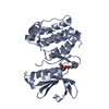
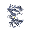
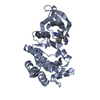
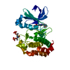
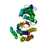
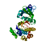
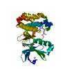
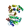
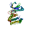
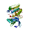
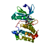
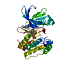
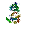
Aurora kinase A / Aurora kinase / Phosphorylase Kinase; domain 1 / Phosphorylase Kinase; domain 1 / Transferase(Phosphotransferase) domain 1 / Transferase(Phosphotransferase); domain 1 / Serine/threonine-protein kinase, active site / Serine/Threonine protein kinases active-site signature. / Protein kinase domain / Serine/Threonine protein kinases, catalytic domain ...Aurora kinase A / Aurora kinase / Phosphorylase Kinase; domain 1 / Phosphorylase Kinase; domain 1 / Transferase(Phosphotransferase) domain 1 / Transferase(Phosphotransferase); domain 1 / Serine/threonine-protein kinase, active site / Serine/Threonine protein kinases active-site signature. / Protein kinase domain / Serine/Threonine protein kinases, catalytic domain / Protein kinase, ATP binding site / Protein kinases ATP-binding region signature. / Protein kinase domain profile. / Protein kinase domain / Protein kinase-like domain superfamily / 2-Layer Sandwich / Orthogonal Bundle / Mainly Alpha / Alpha Beta Similarity search - Domain/homology
In the structure databanks used in Yorodumi, some data are registered as the other names, "COVID-19 virus" and "2019-nCoV". Here are the details of the virus and the list of structure data.
Jan 31, 2019. EMDB accession codes are about to change! (news from PDBe EMDB page)
EMDB accession codes are about to change! (news from PDBe EMDB page)
The allocation of 4 digits for EMDB accession codes will soon come to an end. Whilst these codes will remain in use, new EMDB accession codes will include an additional digit and will expand incrementally as the available range of codes is exhausted. The current 4-digit format prefixed with “EMD-” (i.e. EMD-XXXX) will advance to a 5-digit format (i.e. EMD-XXXXX), and so on. It is currently estimated that the 4-digit codes will be depleted around Spring 2019, at which point the 5-digit format will come into force.
The EM Navigator/Yorodumi systems omit the EMD- prefix.
Related info.:Q: What is EMD? / ID/Accession-code notation in Yorodumi/EM Navigator
Yorodumi is a browser for structure data from EMDB, PDB, SASBDB, etc.
This page is also the successor to EM Navigator detail page, and also detail information page/front-end page for Omokage search.
The word "yorodu" (or yorozu) is an old Japanese word meaning "ten thousand". "mi" (miru) is to see.
Related info.:EMDB / PDB / SASBDB / Comparison of 3 databanks / Yorodumi Search / Aug 31, 2016. New EM Navigator & Yorodumi / Yorodumi Papers / Jmol/JSmol / Function and homology information / Changes in new EM Navigator and Yorodumi
 Movie
Movie Controller

 Yorodumi
 Open data
 Basic information
 Components
 Keywords
 Function and homology information
 HOMO SAPIENS (human)
 X-RAY DIFFRACTION /
  Authors
 Citation
 Structure visualization
 Downloads & links
 Other downloads
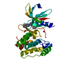


 PDBj
PDBj


 Assembly
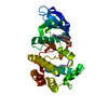


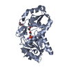
 Mass: 513.012 Da / Num. of mol.: 1 / Source method: obtained synthetically / Formula: C24H25ClN6O3S
 Mass: 18.015 Da / Num. of mol.: 25 / Source method: isolated from a natural source / Formula: H2O
 Sample preparation
 / Beamline: ID14-1 / Wavelength: 0.9334
  Processing