| Entry | Database: PDB / ID: 2wm3
|
|---|
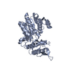
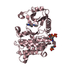
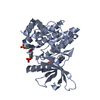
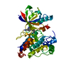
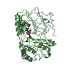
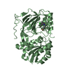
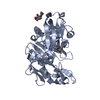
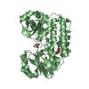
| Title | Crystal structure of NmrA-like family domain containing protein 1 in complex with niflumic acid |
|---|
 Components Components | NMRA-LIKE FAMILY DOMAIN CONTAINING PROTEIN 1 |
|---|
 Keywords Keywords | PROTEIN BINDING |
|---|
| Function / homology |  Function and homology information Function and homology information
: / NmrA-like domain / NmrA-like family / UDP-galactose 4-epimerase, domain 1 / UDP-galactose 4-epimerase; domain 1 / NAD(P)-binding Rossmann-like Domain / NAD(P)-binding domain superfamily / Alpha-Beta Complex / Rossmann fold / 3-Layer(aba) Sandwich / Alpha BetaSimilarity search - Domain/homology NADP NICOTINAMIDE-ADENINE-DINUCLEOTIDE PHOSPHATE / Chem-NFL / PHOSPHATE ION / NmrA-like family domain-containing protein 1Similarity search - Component |
|---|
| Biological species |  HOMO SAPIENS (human) HOMO SAPIENS (human) |
|---|
| Method |  X-RAY DIFFRACTION / SYNCHROTRON / MOLECULAR REPLACEMENT / Resolution: 1.85 Å X-RAY DIFFRACTION / SYNCHROTRON / MOLECULAR REPLACEMENT / Resolution: 1.85 Å |
|---|
 Authors Authors | Bhatia, C. / Yue, W.W. / Niesen, F. / Pilka, E. / Ugochukwu, E. / Savitsky, P. / Hozjan, V. / Roos, A.K. / Filippakopoulos, P. / von Delft, F. ...Bhatia, C. / Yue, W.W. / Niesen, F. / Pilka, E. / Ugochukwu, E. / Savitsky, P. / Hozjan, V. / Roos, A.K. / Filippakopoulos, P. / von Delft, F. / Heightman, T. / Arrowsmith, C. / Weigelt, J. / Edwards, A. / Bountra, C. / Oppermann, U. |
|---|
 Citation Citation | Journal: To be Published
Title: Crystal Structure of Nmra-Like Family Domain Containing Protein 1 in Complex with Niflumic Acid
Authors: Bhatia, C. / Yue, W.W. / Pilka, E. / Ugochukwu, E. / Savitsky, P. / Hozjan, V. / Roos, A.K. / Filippakopoulos, P. / von Delft, F. / Heightman, T. / Arrowsmith, C. / Weigelt, J. / Edwards, A. ...Authors: Bhatia, C. / Yue, W.W. / Pilka, E. / Ugochukwu, E. / Savitsky, P. / Hozjan, V. / Roos, A.K. / Filippakopoulos, P. / von Delft, F. / Heightman, T. / Arrowsmith, C. / Weigelt, J. / Edwards, A. / Bountra, C. / Oppermann, U. |
|---|
| History | | Deposition | Jun 29, 2009 | Deposition site: PDBE / Processing site: PDBE |
|---|
| Revision 1.0 | Aug 4, 2009 | Provider: repository / Type: Initial release |
|---|
| Revision 1.1 | Jul 13, 2011 | Group: Advisory / Version format compliance |
|---|
| Revision 1.2 | Dec 5, 2012 | Group: Database references / Non-polymer description ...Database references / Non-polymer description / Other / Source and taxonomy / Structure summary |
|---|
| Revision 1.3 | Jan 24, 2018 | Group: Database references / Category: citation_author / Item: _citation_author.name |
|---|
| Revision 1.4 | May 8, 2024 | Group: Data collection / Database references ...Data collection / Database references / Derived calculations / Other
Category: chem_comp_atom / chem_comp_bond ...chem_comp_atom / chem_comp_bond / database_2 / pdbx_database_status / struct_site
Item: _database_2.pdbx_DOI / _database_2.pdbx_database_accession ..._database_2.pdbx_DOI / _database_2.pdbx_database_accession / _pdbx_database_status.status_code_sf / _struct_site.pdbx_auth_asym_id / _struct_site.pdbx_auth_comp_id / _struct_site.pdbx_auth_seq_id |
|---|
|
|---|
 Movie
Movie Controller

 Yorodumi
 Open data
 Basic information
 Structure visualization
 Downloads & links
 Other downloads
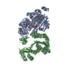


 PDBj
PDBj
 Assembly


 Mass: 743.405 Da / Num. of mol.: 1 / Source method: obtained synthetically / Formula: C21H28N7O17P3
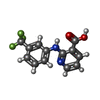
 Mass: 282.218 Da / Num. of mol.: 2 / Source method: obtained synthetically / Formula: C13H9F3N2O2 / Comment: inhibitor*YM
 Mass: 94.971 Da / Num. of mol.: 1 / Source method: obtained synthetically / Formula: PO4
 Mass: 92.094 Da / Num. of mol.: 1 / Source method: obtained synthetically / Formula: C3H8O3
 Mass: 35.453 Da / Num. of mol.: 1 / Source method: obtained synthetically / Formula: Cl
 Sample preparation
 / Beamline: X10SA / Wavelength: 0.9764
  Processing