 Movie
Movie Controller

[English] 日本語
 Yorodumi
- PDB-2vq4: Carbohydrate-binding of the starch binding domain of Rhizopus ory... -
+ Open data

- Basic information
| Entry | Database: PDB / ID: 2vq4 | ||||||
|---|---|---|---|---|---|---|---|
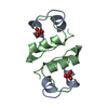
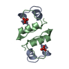
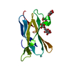
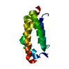
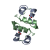
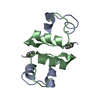
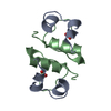
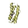
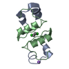
| Title | Carbohydrate-binding of the starch binding domain of Rhizopus oryzae glucoamylase in complex with beta-cyclodextrin and maltoheptaose | ||||||
 Components Components | GLUCOAMYLASE A | ||||||
 Keywords Keywords |  HYDROLASE / RHIZOPUS ORYZAE GLUCOAMYLASE / CARBOHYDRATE BINDING / STARCH BINDING DOMAIN HYDROLASE / RHIZOPUS ORYZAE GLUCOAMYLASE / CARBOHYDRATE BINDING / STARCH BINDING DOMAIN | ||||||
| Function / homology |  Function and homology information Function and homology informationpolysaccharide metabolic process / glucan 1,4-alpha-glucosidase / glucan 1,4-alpha-glucosidase activity Similarity search - Function | ||||||
| Biological species |  RHIZOPUS ORYZAE (fungus) RHIZOPUS ORYZAE (fungus) | ||||||
| Method | X-RAY DIFFRACTION / SYNCHROTRON / MOLECULAR REPLACEMENT / Resolution: 1.25 Å | ||||||
 Authors Authors | Tung, J.-Y. / Liu, Y.-Y. / Sun, Y.-J. | ||||||
 Citation Citation | Journal: Biochem.J. / Year: 2008 Title: Crystal Structures of the Starch-Binding Domain from Rhizopus Oryzae Glucoamylase Reveal a Polysaccharide-Binding Path. Authors: Tung, J.-Y. / Chang, M.D. / Chou, W. / Liu, Y.-Y. / Yeh, Y. / Chang, F. / Lin, S. / Qiu, Z. / Sun, Y.-J. | ||||||
| History |
| ||||||
| Remark 700 | SHEET THE SHEET STRUCTURE OF THIS MOLECULE IS BIFURCATED. IN ORDER TO REPRESENT THIS FEATURE IN ... SHEET THE SHEET STRUCTURE OF THIS MOLECULE IS BIFURCATED. IN ORDER TO REPRESENT THIS FEATURE IN THE SHEET RECORDS BELOW, TWO SHEETS ARE DEFINED. |
- Structure visualization
| Structure viewer | Molecule: MolmilJmol/JSmol |
|---|
- Downloads & links
-Download
| PDBx/mmCIF format | 2vq4.cif.gz | 62.1 KB | Display | PDBx/mmCIF format |
|---|---|---|---|---|
| PDB format | pdb2vq4.ent.gz | 45.7 KB | Display | PDB format |
| PDBx/mmJSON format | 2vq4.json.gz | Tree view | PDBx/mmJSON format | |
| Others |  Other downloads Other downloads |
-Validation report
| Arichive directory | https://data.pdbj.org/pub/pdb/validation_reports/vq/2vq4ftp://data.pdbj.org/pub/pdb/validation_reports/vq/2vq4 | HTTPS FTP |
|---|
-Related structure data
| Related structure data |  2v8lSC  2v8mC S: Starting model for refinement C: citing same article ( |
|---|---|
| Similar structure data |

-Links
 PDBj
PDBj
- Assembly
| Deposited unit | 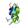
| ||||||||
|---|---|---|---|---|---|---|---|---|---|
| 1 |
| ||||||||
| Unit cell |
|
-Components
| #1: Protein | Mass: 11662.581 Da / Num. of mol.: 1 / Fragment: STARCH BINDING DOMAIN, RESIDUES 26-131 / Mutation: YES Source method: isolated from a genetically manipulated source Details: ILE 53 IS A CLONING VARIANT FROM A LOCAL STRAIN OF R. ORYZAE Source: (gene. exp.) RHIZOPUS ORYZAE (fungus) / Plasmid: PET23A / Production host:  ESCHERICHIA COLI (E. coli) / Strain (production host): BL21 (DE3) / References: UniProt: Q2VC81, glucan 1,4-alpha-glucosidase ESCHERICHIA COLI (E. coli) / Strain (production host): BL21 (DE3) / References: UniProt: Q2VC81, glucan 1,4-alpha-glucosidase | ||
|---|---|---|---|
| #2: Water | ChemComp-HOH / Water Mass: 18.015 Da / Num. of mol.: 210 / Source method: isolated from a natural source / Formula: H2O Mass: 18.015 Da / Num. of mol.: 210 / Source method: isolated from a natural source / Formula: H2O | ||
| Compound details | ENGINEERED| Sequence details | ILE 53 IS A CLONING VARIANT FROM A LOCAL STRAIN OF R. ORYZAE | |
-Experimental details
-Experiment
| Experiment | Method: X-RAY DIFFRACTION |
|---|
- Sample preparation
| Crystal | Density Matthews: 1.81 Å3/Da / Density % sol: 0.32 % / Description: NONE |
|---|---|
| Crystal grow | Details: 25% PEG4000 |
-Data collection
| Diffraction | Mean temperature: 273 K |
|---|---|
| Diffraction source | Source: SYNCHROTRON / Site: NSRRC  / Beamline: BL13C1 / Wavelength: 0.9732 / Beamline: BL13C1 / Wavelength: 0.9732 |
| Detector | Type: ADSC CCD / Detector: CCD / Date: Feb 1, 2008 |
| Radiation | Protocol: SINGLE WAVELENGTH / Monochromatic (M) / Laue (L): M / Scattering type: x-ray |
| Radiation wavelength | Wavelength: 0.9732 Å / Relative weight: 1 |
| Reflection | Resolution: 1.25→30 Å / Num. obs: 23978 / % possible obs: 98.5 % / Observed criterion σ(I): 2 / Redundancy: 11.6 % / Biso Wilson estimate: 16 Å2 / Rmerge(I) obs: 0.07 / Net I/σ(I): 31.5 |
| Reflection shell | Resolution: 1.25→1.29 Å / Redundancy: 10.3 % / Rmerge(I) obs: 0.29 / Mean I/σ(I) obs: 6.7 / % possible all: 93.9 |
- Processing
| Software |
| ||||||||||||||||||||||||||||||||||||||||||||||||||||||||||||||||||||||||||||||||||||||||||||||||||||||||||||||||||||||||||||||||||||||||||||||||||||||||||||||||||||||||||||||||||||||
|---|---|---|---|---|---|---|---|---|---|---|---|---|---|---|---|---|---|---|---|---|---|---|---|---|---|---|---|---|---|---|---|---|---|---|---|---|---|---|---|---|---|---|---|---|---|---|---|---|---|---|---|---|---|---|---|---|---|---|---|---|---|---|---|---|---|---|---|---|---|---|---|---|---|---|---|---|---|---|---|---|---|---|---|---|---|---|---|---|---|---|---|---|---|---|---|---|---|---|---|---|---|---|---|---|---|---|---|---|---|---|---|---|---|---|---|---|---|---|---|---|---|---|---|---|---|---|---|---|---|---|---|---|---|---|---|---|---|---|---|---|---|---|---|---|---|---|---|---|---|---|---|---|---|---|---|---|---|---|---|---|---|---|---|---|---|---|---|---|---|---|---|---|---|---|---|---|---|---|---|---|---|---|---|
| Refinement | Method to determine structure: MOLECULAR REPLACEMENT Starting model: PDB ENTRY 2V8L Resolution: 1.25→30 Å / Cor.coef. Fo:Fc: 0.973 / Cor.coef. Fo:Fc free: 0.968 / SU B: 1.288 / SU ML: 0.026 / Cross valid method: THROUGHOUT / σ(F): 2 / ESU R: 0.053 / ESU R Free: 0.047 / Stereochemistry target values: MAXIMUM LIKELIHOOD Details: HYDROGENS HAVE BEEN ADDED IN THE RIDING POSITIONS. RESIDUES 6, 42, 99, AND 101 ARE DISORDERED ALTERNATE POSITIONS ARE PRESENT FOR SIDE CHAIN OF RESIDUES SER 8, GLN10, SER 19, THR37, SER 44, ...Details: HYDROGENS HAVE BEEN ADDED IN THE RIDING POSITIONS. RESIDUES 6, 42, 99, AND 101 ARE DISORDERED ALTERNATE POSITIONS ARE PRESENT FOR SIDE CHAIN OF RESIDUES SER 8, GLN10, SER 19, THR37, SER 44, SER57, GLU68, AND SER89. HENCE THEIR ALTERNATE INDICATORS ARE A AND B. THEY CONCERNED HAVE OCCUPANCY BETWEEN 0.0 AND 1.0 AND WERE REFINED AS DESCRIBED ABOVE. OCCUPANCY VALUES LOWER THAN 1. 0 APPEARED TO JUSTIFY BETTER THE ELECTRON DENSITY.
| ||||||||||||||||||||||||||||||||||||||||||||||||||||||||||||||||||||||||||||||||||||||||||||||||||||||||||||||||||||||||||||||||||||||||||||||||||||||||||||||||||||||||||||||||||||||
| Solvent computation | Ion probe radii: 0.8 Å / Shrinkage radii: 0.8 Å / VDW probe radii: 1.2 Å / Solvent model: BABINET MODEL WITH MASK | ||||||||||||||||||||||||||||||||||||||||||||||||||||||||||||||||||||||||||||||||||||||||||||||||||||||||||||||||||||||||||||||||||||||||||||||||||||||||||||||||||||||||||||||||||||||
| Displacement parameters | Biso mean: 12.06 Å2
| ||||||||||||||||||||||||||||||||||||||||||||||||||||||||||||||||||||||||||||||||||||||||||||||||||||||||||||||||||||||||||||||||||||||||||||||||||||||||||||||||||||||||||||||||||||||
| Refinement step | Cycle: LAST / Resolution: 1.25→30 Å
| ||||||||||||||||||||||||||||||||||||||||||||||||||||||||||||||||||||||||||||||||||||||||||||||||||||||||||||||||||||||||||||||||||||||||||||||||||||||||||||||||||||||||||||||||||||||
| Refine LS restraints |
|