 Movie
Movie Controller

[English] 日本語
 Yorodumi
- PDB-2r66: Complex Structure of Sucrose Phosphate Synthase (SPS)-F6P of Halo... -
+ Open data

- Basic information
| Entry | Database: PDB / ID: 2r66 | ||||||
|---|---|---|---|---|---|---|---|
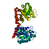
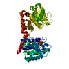
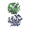
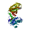
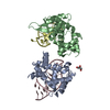
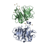
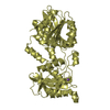
| Title | Complex Structure of Sucrose Phosphate Synthase (SPS)-F6P of Halothermothrix orenii | ||||||
 Components Components | Glycosyl transferase, group 1 Glycosyltransferase Glycosyltransferase | ||||||
 Keywords Keywords | TRANSFERASE / Rossmann-fold | ||||||
| Function / homology |  Function and homology informationsucrose-phosphate synthase / sucrose-phosphate synthase activity / sucrose synthase activity / sucrose metabolic process Function and homology informationsucrose-phosphate synthase / sucrose-phosphate synthase activity / sucrose synthase activity / sucrose metabolic processSimilarity search - Function | ||||||
| Biological species |  Halothermothrix orenii (bacteria) Halothermothrix orenii (bacteria) | ||||||
| Method | X-RAY DIFFRACTION / MOLECULAR REPLACEMENT / Resolution: 2.8 Å | ||||||
 Authors Authors | Sivaraman, J. / Chua, T.K. | ||||||
 Citation Citation | Journal: Plant Cell / Year: 2008 Title: The Structure of Sucrose Phosphate Synthase from Halothermothrix orenii Reveals Its Mechanism of Action and Binding Mode Authors: Chua, T.K. / Bujnicki, J.M. / Tan, T.-C. / Huynh, F. / Patel, B.K. / Sivaraman, J. | ||||||
| History |
|
- Structure visualization
| Structure viewer | Molecule: MolmilJmol/JSmol |
|---|
- Downloads & links
-Download
| PDBx/mmCIF format | 2r66.cif.gz | 107.2 KB | Display | PDBx/mmCIF format |
|---|---|---|---|---|
| PDB format | pdb2r66.ent.gz | 80.9 KB | Display | PDB format |
| PDBx/mmJSON format | 2r66.json.gz | Tree view | PDBx/mmJSON format | |
| Others |  Other downloads Other downloads |
-Validation report
| Arichive directory | https://data.pdbj.org/pub/pdb/validation_reports/r6/2r66ftp://data.pdbj.org/pub/pdb/validation_reports/r6/2r66 | HTTPS FTP |
|---|
-Related structure data
| Related structure data |  2r60SC  2r68C C: citing same article ( S: Starting model for refinement |
|---|---|
| Similar structure data |


-Links
 PDBj
PDBj

- Assembly
| Deposited unit | 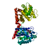
| ||||||||
|---|---|---|---|---|---|---|---|---|---|
| 1 |
| ||||||||
| Unit cell |
|
-Components
| #1: Protein | Glycosyltransferase / Sucrose Phosphate Synthase Mass: 56882.742 Da / Num. of mol.: 1 Source method: isolated from a genetically manipulated source Source: (gene. exp.) Halothermothrix orenii (bacteria) / Strain: H 168 / Gene: SPS / Plasmid: pTrcHisA / Production host: Escherichia coli (E. coli) / Strain (production host): BL21(DE3)References: UniProt: Q2ADF5, UniProt: B8CZ51*PLUS, sucrose-phosphate synthase |
|---|---|
| #2: Sugar | ChemComp-F6P / Fructose 6-phosphate  Type: D-saccharide, beta linking / Mass: 260.136 Da / Num. of mol.: 1 Type: D-saccharide, beta linking / Mass: 260.136 Da / Num. of mol.: 1Source method: isolated from a genetically manipulated source Formula: C6H13O9P |
| #3: Water | ChemComp-HOH / Water Mass: 18.015 Da / Num. of mol.: 118 / Source method: isolated from a natural source / Formula: H2O Mass: 18.015 Da / Num. of mol.: 118 / Source method: isolated from a natural source / Formula: H2O |
| Sequence details | THE FIRST THREE RESIDUES, MET 1, VAL 2, AND GLU 3 ARE PART OF THE GENE PRODUCT. |
-Experimental details
-Experiment
| Experiment | Method: X-RAY DIFFRACTION / Number of used crystals: 1 |
|---|
- Sample preparation
| Crystal | Density Matthews: 2.42 Å3/Da / Density % sol: 49.23 % |
|---|---|
| Crystal grow | Temperature: 298 K / Method: vapor diffusion, hanging drop / pH: 6.5 Details: 20% PEG 4000, 0.6M NaCl, 0.1M Na MES, pH 6.5, VAPOR DIFFUSION, HANGING DROP, temperature 298K |
-Data collection
| Diffraction | Mean temperature: 100 K | ||||||||||||||||||||||||||||||||||||||||||||||||||||||||||||||||||
|---|---|---|---|---|---|---|---|---|---|---|---|---|---|---|---|---|---|---|---|---|---|---|---|---|---|---|---|---|---|---|---|---|---|---|---|---|---|---|---|---|---|---|---|---|---|---|---|---|---|---|---|---|---|---|---|---|---|---|---|---|---|---|---|---|---|---|---|
| Diffraction source | Source: ROTATING ANODE / Type: RIGAKU / Wavelength: 1.5418 Å | ||||||||||||||||||||||||||||||||||||||||||||||||||||||||||||||||||
| Detector | Type: RIGAKU RAXIS IV++ / Detector: IMAGE PLATE / Date: Jun 13, 2006 | ||||||||||||||||||||||||||||||||||||||||||||||||||||||||||||||||||
| Radiation | Protocol: SINGLE WAVELENGTH / Monochromatic (M) / Laue (L): M / Scattering type: x-ray | ||||||||||||||||||||||||||||||||||||||||||||||||||||||||||||||||||
| Radiation wavelength | Wavelength: 1.5418 Å / Relative weight: 1 | ||||||||||||||||||||||||||||||||||||||||||||||||||||||||||||||||||
| Reflection | Resolution: 2.8→50 Å / Num. obs: 13734 / % possible obs: 99.8 % / Redundancy: 3.7 % / Rmerge(I) obs: 0.075 / Χ2: 1.394 / Net I/σ(I): 14.6 | ||||||||||||||||||||||||||||||||||||||||||||||||||||||||||||||||||
| Reflection shell |
|
- Processing
| Software |
| ||||||||||||||||||||||||||||||||||||
|---|---|---|---|---|---|---|---|---|---|---|---|---|---|---|---|---|---|---|---|---|---|---|---|---|---|---|---|---|---|---|---|---|---|---|---|---|---|
| Refinement | Method to determine structure: MOLECULAR REPLACEMENT Starting model: PDB ENTRY 2R60 Resolution: 2.8→20 Å / Cross valid method: THROUGHOUT / σ(F): 2
| ||||||||||||||||||||||||||||||||||||
| Solvent computation | Bsol: 52.904 Å2 | ||||||||||||||||||||||||||||||||||||
| Displacement parameters | Biso mean: 46.807 Å2
| ||||||||||||||||||||||||||||||||||||
| Refinement step | Cycle: LAST / Resolution: 2.8→20 Å
| ||||||||||||||||||||||||||||||||||||
| Refine LS restraints |
| ||||||||||||||||||||||||||||||||||||
| Xplor file |
|
