 Movie
Movie Controller


+ Open data

- Basic information
| Entry | Database: PDB / ID: 2qw1 | ||||||
|---|---|---|---|---|---|---|---|
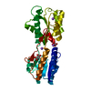
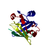
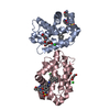
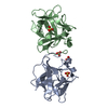
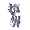
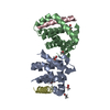
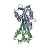
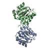
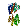
| Title | Glucose/galactose binding protein bound to 3-O-methyl D-glucose | ||||||
 Components Components | D-galactose-binding periplasmic protein | ||||||
 Keywords Keywords |  TRANSPORT PROTEIN / PERIPLASMIC BINDING PROTEIN / ANTAGONIST / CHEMOTAXIS / TRANSPORT / GGBP / 3-O-METHYL GLUCOSE / Sugar transport TRANSPORT PROTEIN / PERIPLASMIC BINDING PROTEIN / ANTAGONIST / CHEMOTAXIS / TRANSPORT / GGBP / 3-O-METHYL GLUCOSE / Sugar transport | ||||||
| Function / homology |  Function and homology information Function and homology informationmethylgalactoside transport / galactose transmembrane transport / ATP-binding cassette (ABC) transporter complex, substrate-binding subunit-containing / chemotaxis / outer membrane-bounded periplasmic space / carbohydrate binding / calcium ion binding / membraneSimilarity search - Function | ||||||
| Biological species |  Escherichia coli (E. coli) Escherichia coli (E. coli) | ||||||
| Method | X-RAY DIFFRACTION / MOLECULAR REPLACEMENT / Resolution: 1.7 Å | ||||||
 Authors Authors | Borrok, M.J. / Kiessling, L.L. / Forest, K.T. | ||||||
 Citation Citation | Journal: Acs Chem.Biol. / Year: 2009 Title: Structure-based design of a periplasmic binding protein antagonist that prevents domain closure. Authors: Borrok, M.J. / Zhu, Y. / Forest, K.T. / Kiessling, L.L. | ||||||
| History |
|
- Structure visualization
| Structure viewer | Molecule: MolmilJmol/JSmol |
|---|
- Downloads & links
-Download
| PDBx/mmCIF format | 2qw1.cif.gz | 82.5 KB | Display | PDBx/mmCIF format |
|---|---|---|---|---|
| PDB format | pdb2qw1.ent.gz | 59.8 KB | Display | PDB format |
| PDBx/mmJSON format | 2qw1.json.gz | Tree view | PDBx/mmJSON format | |
| Others |  Other downloads Other downloads |
-Validation report
| Arichive directory | https://data.pdbj.org/pub/pdb/validation_reports/qw/2qw1ftp://data.pdbj.org/pub/pdb/validation_reports/qw/2qw1 | HTTPS FTP |
|---|
-Related structure data
| Related structure data |  2fw0S S: Starting model for refinement |
|---|---|
| Similar structure data |


-Links
 PDBj
PDBj- Assembly
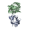
| Deposited unit | 
| ||||||||
|---|---|---|---|---|---|---|---|---|---|
| 1 |
| ||||||||
| Unit cell |
|
-Components
| #1: Protein | Mass: 33407.625 Da / Num. of mol.: 1 Source method: isolated from a genetically manipulated source Source: (gene. exp.) Escherichia coli (E. coli) / Strain: K12 / Gene: mglB / Plasmid: pVB2 / Production host: Escherichia coli (E. coli) / Strain (production host): HB929 / References: UniProt: P0AEE5 | ||
|---|---|---|---|
| #2: Sugar | ChemComp-3MG /   Type: D-saccharide, beta linking / Mass: 194.182 Da / Num. of mol.: 1 Type: D-saccharide, beta linking / Mass: 194.182 Da / Num. of mol.: 1Source method: isolated from a genetically manipulated source Formula: C7H14O6 | ||
| #3: Chemical | ChemComp-CA /   Mass: 40.078 Da / Num. of mol.: 1 / Source method: obtained synthetically / Formula: Ca Mass: 40.078 Da / Num. of mol.: 1 / Source method: obtained synthetically / Formula: Ca | ||
| #4: Chemical |   Mass: 22.990 Da / Num. of mol.: 2 / Source method: obtained synthetically / Formula: Na Mass: 22.990 Da / Num. of mol.: 2 / Source method: obtained synthetically / Formula: Na#5: Water | ChemComp-HOH / | Water Mass: 18.015 Da / Num. of mol.: 351 / Source method: isolated from a natural source / Formula: H2O Mass: 18.015 Da / Num. of mol.: 351 / Source method: isolated from a natural source / Formula: H2O |
-Experimental details
-Experiment
| Experiment | Method: X-RAY DIFFRACTION / Number of used crystals: 1 |
|---|
- Sample preparation
| Crystal | Density Matthews: 3.5 Å3/Da / Density % sol: 64.87 % |
|---|---|
| Crystal grow | Temperature: 298 K / Method: vapor diffusion, hanging drop / pH: 6 Details: 2.0 M ammonium sulfate and 0.05 M sodium citrate dehydrate, 3-O-methyl glucose (5mM) was soaked in. 2.5 M Na Malonate cryoprotectant, pH 6.0, VAPOR DIFFUSION, HANGING DROP, temperature 298K |
-Data collection
| Diffraction | Mean temperature: 100 K |
|---|---|
| Diffraction source | Source: ROTATING ANODE / Type: BRUKER AXS MICROSTAR / Wavelength: 1.54 Å |
| Detector | Type: Bruker Platinum 135 / Detector: CCD / Date: Jul 25, 2005 |
| Radiation | Monochromator: GLOEBEL MIRRORS / Protocol: SINGLE WAVELENGTH / Monochromatic (M) / Laue (L): M / Scattering type: x-ray |
| Radiation wavelength | Wavelength: 1.54 Å / Relative weight: 1 |
| Reflection | Resolution: 1.7→50 Å / Num. all: 52428 / Num. obs: 47220 / % possible obs: 90.1 % / Observed criterion σ(F): 0 / Observed criterion σ(I): 0 / Redundancy: 6.31 % / Rsym value: 0.0341 / Net I/σ(I): 25.15 |
| Reflection shell | Resolution: 1.7→1.75 Å / Redundancy: 2.11 % / Mean I/σ(I) obs: 3.82 / Num. unique all: 1972 / Rsym value: 0.2589 / % possible all: 56.8 |
- Processing
| Software |
| |||||||||||||||||||||||||||||||||||||||||||||||||||||||||||||||||||||||||||||||||||||||||||||||
|---|---|---|---|---|---|---|---|---|---|---|---|---|---|---|---|---|---|---|---|---|---|---|---|---|---|---|---|---|---|---|---|---|---|---|---|---|---|---|---|---|---|---|---|---|---|---|---|---|---|---|---|---|---|---|---|---|---|---|---|---|---|---|---|---|---|---|---|---|---|---|---|---|---|---|---|---|---|---|---|---|---|---|---|---|---|---|---|---|---|---|---|---|---|---|---|---|
| Refinement | Method to determine structure: MOLECULAR REPLACEMENT Starting model: 2FW0 Resolution: 1.7→40 Å / Cor.coef. Fo:Fc: 0.951 / Cor.coef. Fo:Fc free: 0.944 / Cross valid method: THROUGHOUT / σ(F): 0 / σ(I): 0 / ESU R: 0.094 / ESU R Free: 0.092 / Stereochemistry target values: MAXIMUM LIKELIHOOD / Details: HYDROGENS HAVE BEEN ADDED IN THE RIDING POSITIONS
| |||||||||||||||||||||||||||||||||||||||||||||||||||||||||||||||||||||||||||||||||||||||||||||||
| Solvent computation | Ion probe radii: 0.8 Å / Shrinkage radii: 0.8 Å / VDW probe radii: 1.2 Å / Solvent model: MASK | |||||||||||||||||||||||||||||||||||||||||||||||||||||||||||||||||||||||||||||||||||||||||||||||
| Displacement parameters | Biso mean: 13.533 Å2
| |||||||||||||||||||||||||||||||||||||||||||||||||||||||||||||||||||||||||||||||||||||||||||||||
| Refine analyze | Luzzati coordinate error obs: 0.1874 Å | |||||||||||||||||||||||||||||||||||||||||||||||||||||||||||||||||||||||||||||||||||||||||||||||
| Refinement step | Cycle: LAST / Resolution: 1.7→40 Å
| |||||||||||||||||||||||||||||||||||||||||||||||||||||||||||||||||||||||||||||||||||||||||||||||
| Refine LS restraints |
| |||||||||||||||||||||||||||||||||||||||||||||||||||||||||||||||||||||||||||||||||||||||||||||||
| LS refinement shell | Resolution: 1.7→1.744 Å / Total num. of bins used: 20
|
