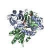
 Movie
Movie Controller


+ Open data

- Basic information
| Entry | Database: PDB / ID: 2qkt | ||||||
|---|---|---|---|---|---|---|---|
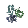
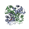
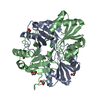
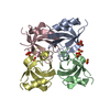
| Title | Crystal Structure of the 5th PDZ domain of InaD | ||||||
 Components Components | Inactivation-no-after-potential D protein | ||||||
 Keywords Keywords | PEPTIDE BINDING PROTEIN / PDZ DOMAIN / SCAFFOLDING PROTEIN / DISULFIDE-BOND / Membrane / Sensory transduction / Vision / SIGNALING PROTEIN | ||||||
| Function / homology |  Function and homology information Function and homology informationmyosin III binding / detection of light stimulus involved in sensory perception / inaD signaling complex / negative regulation of opsin-mediated signaling pathway / Antigen processing: Ubiquitination & Proteasome degradation / rhabdomere / cellular response to light stimulus / myosin binding / photoreceptor activity / phototransduction ...myosin III binding / detection of light stimulus involved in sensory perception / inaD signaling complex / negative regulation of opsin-mediated signaling pathway / Antigen processing: Ubiquitination & Proteasome degradation / rhabdomere / cellular response to light stimulus / myosin binding / photoreceptor activity / phototransduction / visual perception / sensory perception of sound / protein localization / signaling receptor complex adaptor activity / calmodulin binding Similarity search - Function | ||||||
| Biological species |  | ||||||
| Method |  X-RAY DIFFRACTION / MIR / Resolution: 2.05 Å X-RAY DIFFRACTION / MIR / Resolution: 2.05 Å | ||||||
 Authors Authors | Ranganathan, R. / Socolich, M. | ||||||
 Citation Citation | Journal: Cell(Cambridge,Mass.) / Year: 2007 Title: Dynamic Scaffolding in a G Protein-Coupled Signaling System. Authors: Mishra, P. / Socolich, M. / Wall, M.A. / Graves, J. / Wang, Z. / Ranganathan, R. | ||||||
| History |
|
- Structure visualization
| Structure viewer | Molecule: MolmilJmol/JSmol |
|---|
- Downloads & links
-Download
| PDBx/mmCIF format | 2qkt.cif.gz | 43.9 KB | Display | PDBx/mmCIF format |
|---|---|---|---|---|
| PDB format | pdb2qkt.ent.gz | 34.3 KB | Display | PDB format |
| PDBx/mmJSON format | 2qkt.json.gz | Tree view | PDBx/mmJSON format | |
| Others |  Other downloads Other downloads |
-Validation report
| Summary document | 2qkt_validation.pdf.gz | 428.3 KB | Display | wwPDB validaton report |
|---|---|---|---|---|
| Full document | 2qkt_full_validation.pdf.gz | 430.3 KB | Display | |
| Data in XML | 2qkt_validation.xml.gz | 10.2 KB | Display | |
| Data in CIF | 2qkt_validation.cif.gz | 13.8 KB | Display | |
| Arichive directory | https://data.pdbj.org/pub/pdb/validation_reports/qk/2qktftp://data.pdbj.org/pub/pdb/validation_reports/qk/2qkt | HTTPS FTP |
-Related structure data


-Links
 PDBj
PDBj- Assembly
| Deposited unit | 
| ||||||||
|---|---|---|---|---|---|---|---|---|---|
| 1 | 
| ||||||||
| Unit cell |
| ||||||||
| Details | The biological assembly is unknown. |
-Components
| #1: Protein | Mass: 9711.200 Da / Num. of mol.: 2 / Fragment: 5th PDZ domain Source method: isolated from a genetically manipulated source Source: (gene. exp.)  #2: Water | ChemComp-HOH / |  Mass: 18.015 Da / Num. of mol.: 153 / Source method: isolated from a natural source / Formula: H2O Mass: 18.015 Da / Num. of mol.: 153 / Source method: isolated from a natural source / Formula: H2O |
|---|
-Experimental details
-Experiment
| Experiment | Method: X-RAY DIFFRACTION / Number of used crystals: 4 |
|---|
- Sample preparation
| Crystal | Density Matthews: 2.99 Å3/Da / Density % sol: 58.82 % |
|---|---|
| Crystal grow | Temperature: 277 K / Method: vapor diffusion, hanging drop / pH: 7.5 Details: 1.3M Sodium Citrate, pH 7.5, VAPOR DIFFUSION, HANGING DROP, temperature 277K |
-Data collection
| Diffraction |
| |||||||||||||||||||||||||
|---|---|---|---|---|---|---|---|---|---|---|---|---|---|---|---|---|---|---|---|---|---|---|---|---|---|---|
| Diffraction source |
| |||||||||||||||||||||||||
| Detector |
| |||||||||||||||||||||||||
| Radiation |
| |||||||||||||||||||||||||
| Radiation wavelength | Wavelength: 1.54 Å / Relative weight: 1 | |||||||||||||||||||||||||
| Reflection | Resolution: 2.05→20 Å / Num. obs: 14738 / % possible obs: 99.7 % / Biso Wilson estimate: 15.4 Å2 / Rsym value: 0.044 / Net I/σ(I): 0.443 | |||||||||||||||||||||||||
| Reflection shell | Resolution: 2.05→2.16 Å / Mean I/σ(I) obs: 16.7 / Rsym value: 0.153 / % possible all: 99.8 |
- Processing
| Software |
| ||||||||||||||||||||||||||||
|---|---|---|---|---|---|---|---|---|---|---|---|---|---|---|---|---|---|---|---|---|---|---|---|---|---|---|---|---|---|
| Refinement | Method to determine structure: MIR / Resolution: 2.05→20 Å / Rfactor Rfree error: 0.009 / Data cutoff high absF: 1082751.5 / Data cutoff low absF: 0 / Isotropic thermal model: RESTRAINED / Cross valid method: THROUGHOUT / σ(F): 0 / Details: BULK SOLVENT MODEL USED
| ||||||||||||||||||||||||||||
| Displacement parameters | Biso mean: 27.5 Å2
| ||||||||||||||||||||||||||||
| Refine analyze |
| ||||||||||||||||||||||||||||
| Refinement step | Cycle: LAST / Resolution: 2.05→20 Å
| ||||||||||||||||||||||||||||
| Refine LS restraints |
| ||||||||||||||||||||||||||||
| LS refinement shell | Resolution: 2.05→2.18 Å / Rfactor Rfree error: 0.024 / Total num. of bins used: 6
| ||||||||||||||||||||||||||||
| Xplor file |
|
